The LIG Nanowise SMAL™ (Super-Resolution Microsphere Amplifying Lens) is an optical module designed to achieve lateral resolution far beyond the diffraction limit of conventional microscopy.
To verify its true resolving power, LIG Nanowise evaluated the SMAL lens using the Newport HIGHRES-1 USAF resolution target, which includes features as small as 137 nm – well below the capabilities of standard optical objectives.
This article describes that evaluation, confirming the SMAL lens’s ability to resolve the smallest features on the HIGHRES-1 target. It also compares SMAL’s performance with both a conventional 100× high-NA objective and SEM ground-truth imaging.
The Newport HIGHRES-1 Target
The HIGHRES-1 target consists of a quartz substrate patterned with a 100 nm chromium layer. This allows a high-precision USAF-1951 resolution chart to be formed.
Its smallest features, group 11, element 6, have 137 nm line widths and 137 nm spaces (3649 lp/mm). These are below the diffraction limit for visible-light microscopy. Newport notes that these structures typically require SEM for clear resolution, meaning that the target is ideally suited to the validation of super-resolution imaging performance.
Experimental Setup
SMAL Lens Optical Imaging Setup
This setup featured:
- LIG Nanowise SMAL lens mounted on LIG Nanowise NANORO M microscope
- High-NA No immersion configuration
- Wideband LED illumination
- Scientific CMOS camera capturing images
The SMAL lens was specifically aligned to maximize near-field coupling, allowing imaging to be performed beyond the diffraction limit.
Control Imaging Using a 100× Objective Lens (UV-Enhanced Reference)
A high-quality 100× UV-capable objective (NA ≈ 1.2) was evaluated as a control. This was done using a methodology reported by JMC Scientific Consulting1 for the Newport HIGHRES-2 slide. Though HIGHRES-2 is a separate model, it shares identical USAF feature dimensions with HIGHRES-1.
The objective resolved features in the ~150-200 nm range under 313 nm UV illumination, capturing Group 10 and parts of Group 11. The smallest 137 nm structures (Group 11, Element 6) were at or below the limit of detection, however. This was consistent with the theoretical diffraction limit, even under UV illumination.
This imaging control confirmed the expected performance:
- The 100× objective resolves down to ~150-200 nm
- This objective cannot reliably resolve 137 nm
- This approach serves as a meaningful high-end optical reference but cannot be considered a super-resolution method
SEM Imaging (Ground-Truth Reference)
Scanning electron microscopy was used to image the HIGHRES-1 target, providing definitive ground-truth resolution. It was observed that the SEM clearly resolved all features down to 137 nm line/space, successfully validating the pattern’s physical dimensions.
SEM is the established method for confirming true resolution because the 137 nm elements are below the diffraction limit of visible-light optics. The SEM image, therefore, serves as the performance benchmark, with SMAL and 100× objective images compared against this.
Results
SEM Reference
SEM imaging successfully resolved all groups and elements, including Group 11, Element 6. It produced clear definition of line widths, spacing, and edges, serving as the definitive verification of the target’s features. [Figure 1]
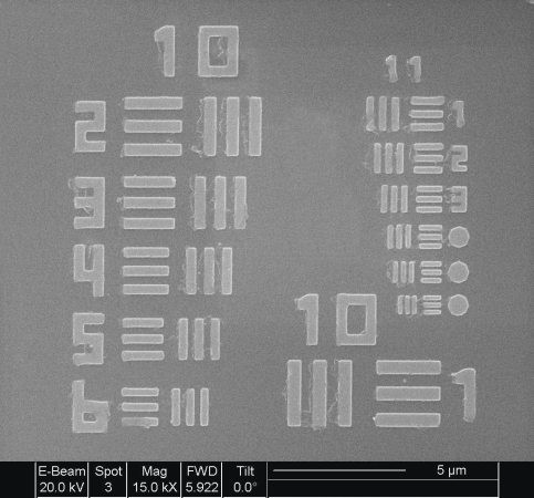
Figure 1. SEM image of the Newport HIGHRES-1 sample target. Image Credit: LIG Nanowise Ltd.
Standard 100× Objective
The 100× objective resolved features down to ~150-200 nm, depending on the wavelength used (Figure 2). This included resolution of Group 10 and partial Group 11 under UV and white light.
The investigation found that 137 nm features remain unresolved, however, with these features appearing blurred or merged. These findings confirm the classical diffraction limit for conventional optics.
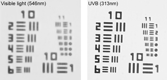
Figure 2. 100x immersion objective lens optical images of the Newport HIGHRES-1 target sample imaged in Visible Light (left) and UV (right). Image Credit: LIG Nanowise Ltd.
SMAL Lens Performance
The SMAL lens was found to clearly resolve the 137 nm features (Group 11, Element 6), as well as capture distinct line/space separation with measurable contrast.
The resolution performance of the lens was found to match the SEM reference closely, demonstrating true super-resolution capability beyond that of the 100× objective.
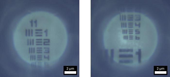
Figure 3. DRY SMAL image of the Newport HIGHRES-1 target sample. Image Credit: LIG Nanowise Ltd.
Comparative Summary
Source: LIG Nanowise Ltd.
| System |
Smallest Resolved
Feature |
Group 11 Element
6(137 nm) |
Notes |
| SEM |
137 nm |
Resolved |
Ground-truth benchmark |
| 100× Objective |
~150–200 nm |
Not resolved |
Diffraction-limited |
| SMAL Lens |
137 nm |
Resolved |
Super-resolution |
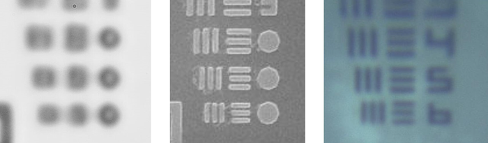
Figure 4. Comparison of the three-imaging method: left: 100x immersion, center: SEM, right: DRY SMAL. Image Credit: LIG Nanowise Ltd.
Conclusion
The LIG Nanowise SMAL lens demonstrated a verified capacity for super-resolution imaging by resolving the smallest 137 nm features of the USAF target, as verified by the Newport HIGHRES-1 target.
The study presented here confirmed that the SMAL lens exhibited comparable performance to SEM and notably superior performance to that of a conventional 100× objective.
These findings highlight the potential of the SMAL lens as a powerful tool for nanoscale imaging in semiconductor inspection, materials science, and nanofabrication quality control.
References and Further Reading
- Crowther, J. (2022). UV Microscopy – Resolution testing with the Newport Highres-2 USAF slide | JMC Scientific Consulting Ltd. (online) JMC Scientific Consulting Ltd. Available at: https://jmcscientificconsulting.com/uv-microscopy-resolution-testing-with-the-newport-highres-2-usaf-slide/.
Acknowledgments
Produced from materials originally authored by LIG Nanowise Limited.

This information has been sourced, reviewed, and adapted from materials provided by LIG Nanowise Ltd.
For more information on this source, please visit LIG Nanowise Ltd.