The Multi-Spheres SMAL uses a novel arrangement of seven microspheres in a honeycomb structure, designed to broaden the lateral field of view while preserving the super-resolution imaging features inherent to SMAL technology.
This setup facilitates concurrent high-resolution imaging across several neighboring areas, enhancing throughput and efficiency for sophisticated microscopy applications.
Product Codes:
MULT-001-BIO-M25-IR → M25 thread, 60 mm par., multisphere, immersion
MULT-001-BIO-RMS-IR → RMS thread, 45 mm par., multisphere immersion
MULT-001-SEM-M25-IR → M25 thread, 60 mm parfocality, multisphere, dry
MULT-001-SEM-RMS-IR → RMS thread, 45 mm parfocality, multisphere, dry
Key Features
- The seven-microsphere honeycomb configuration allows for broader lateral coverage
- Enables simultaneous imaging across various spheres, thereby enhancing throughput
- Preserves sub-diffraction imaging capabilities
- Improves efficiency for surface inspection and material characterization
- Compatible with sophisticated microscope platforms like Nanoro or other optical microscopes from third parties
Intended Applications
- Inspection of microchip and semiconductor surfaces
- Biological imaging with high throughput
- Characterization of advanced materials
- Quality control of surfaces in manufacturing
Specifications
Source: LIG Nanowise Limited
| Attribute |
Description |
| Design |
7-microsphere honeycomb array |
| Immersion |
Oil/water/glycerol/no immersion |
| Resolution |
100 nm |
| Magnification |
180X/200X |
| Integration |
Compatible with Nanoro and other optical microscopes |
| Efficiency |
Increased throughput via multi-zone coverage |
| Thread type |
RMS or M25 x 0.75 mm |
Usage Notes
- Optimal for scenarios that necessitate extensive imaging coverage and elevated throughput
- Most appropriate for surface examination and multi-cellular biological imaging
- The design strikes a balance between complexity and coverage, ensuring wide-area nanoscale imaging
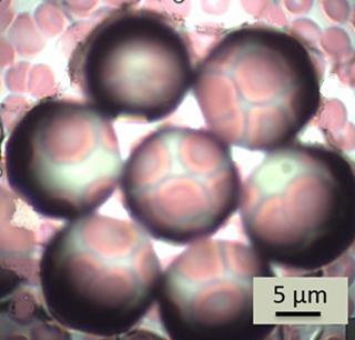
Red blood cells (erythrocytes) imaged with a Multi-Spheres SMAL (Super-resolution Microsphere Amplifying Lens); each microsphere provides a localized super-resolved sub-field. Image Credit: LIG Nanowise Limited