In this interview, industry expert Maria Tesa explains radioluminescence and scintillators, detailing their mechanisms, key performance factors, and how advanced measurement techniques optimize materials for applications in imaging, detection, and high-energy physics.
Please can you introduce radioluminescence and scintillators, and explain their importance?
Maria Tesa: Radioluminescence is the emission of UV, visible, or sometimes near-infrared light from a material when it is excited by high-energy ionizing radiation, such as X-rays, gamma rays, or particles like alpha and beta radiation. Materials that exhibit this behavior are called scintillators, and they can exist as solids, liquids, or gases.
Scintillators are important because they convert ionizing radiation into optical light, which is much easier to detect and quantify using devices like cameras or photomultiplier tubes.
This makes them essential in applications such as medical imaging, airport security scanners, and high-energy physics experiments, including those carried out at large facilities like particle accelerators.
What are the main types of scintillators and how do they differ?
Maria Tesa: There are three main classes of scintillators: inorganic crystals, organic materials, and nanomaterials. Inorganic crystals, such as bismuth germanate or thallium-doped cesium iodide, typically offer high light output but have longer decay times.
Organic scintillators, including materials like anthracene or diphenyl oxazole, tend to have lower light output but much faster response times, which makes them suitable for detecting fast events such as neutron interactions.
More recently, nanomaterials like perovskites and quantum dots have emerged as next-generation scintillators. These materials combine good absorption of high-energy radiation with tunable optical properties, making them very promising for advanced detector technologies.
What are the key properties that define scintillator performance?
Maria Tesa: Some of the most important properties are light output, decay time, and detection efficiency. Light output refers to how effectively the material converts ionizing radiation into optical photons.
Decay time describes how quickly the scintillator emits light after excitation, which determines whether it can resolve fast events.
Other important factors include the limit of detection, linearity, and stability over time. These parameters collectively determine whether a scintillator is suitable for applications like medical imaging, environmental monitoring, or high-energy physics experiments.
Can you explain the mechanism of scintillation in simple terms?
Maria Tesa: In inorganic scintillators, scintillation generally occurs in three stages: conversion, energy transfer, and luminescence.
First, ionizing radiation interacts with the material and generates charge carriers, such as electrons and holes. Then, these charge carriers migrate through the material until they reach luminescence centers, often introduced as dopants or defects.
Finally, when the charge carriers recombine at these centers, they emit a photon in the UV or visible range. This emission is what we detect as scintillation.
What are the key experimental requirements for measuring radioluminescence?
Maria Tesa: To measure radioluminescence, you need a source of ionizing radiation, typically an X-ray source, and a safe enclosure to contain both the source and the sample. The emitted light, which is in the UV, visible, or near-infrared range, can then be directed out of the enclosure into a detection system.
This detection system usually includes a spectrometer capable of both spectral and time-resolved measurements. A photon counting setup is also highly beneficial, as it improves sensitivity and enables accurate quantitative analysis.
How does the XS1 radioluminescence chamber work in practice?
Maria Tesa: The XS1 chamber integrates all the components required for radioluminescence measurements. It contains both continuous and pulsed X-ray sources inside a radiation-safe enclosure.
The emitted light from the sample is collected and transferred via a liquid light guide into a photoluminescence spectrometer, such as the FLS1000 or FS5. This setup allows both steady-state and time-resolved radioluminescence measurements using the same instrument platform.
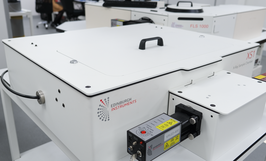
Image Credit: Edinburgh Instruments
Why are both continuous and pulsed X-Ray sources important?
Maria Tesa: The continuous X-ray source is useful for steady-state measurements, such as determining emission spectra, light yield, and stability.
The pulsed X-ray source, on the other hand, enables time-resolved measurements. It is triggered by a pulsed laser, which generates short bursts of X-rays, allowing you to measure radioluminescence lifetimes using techniques like time-correlated single photon counting (TCSPC).
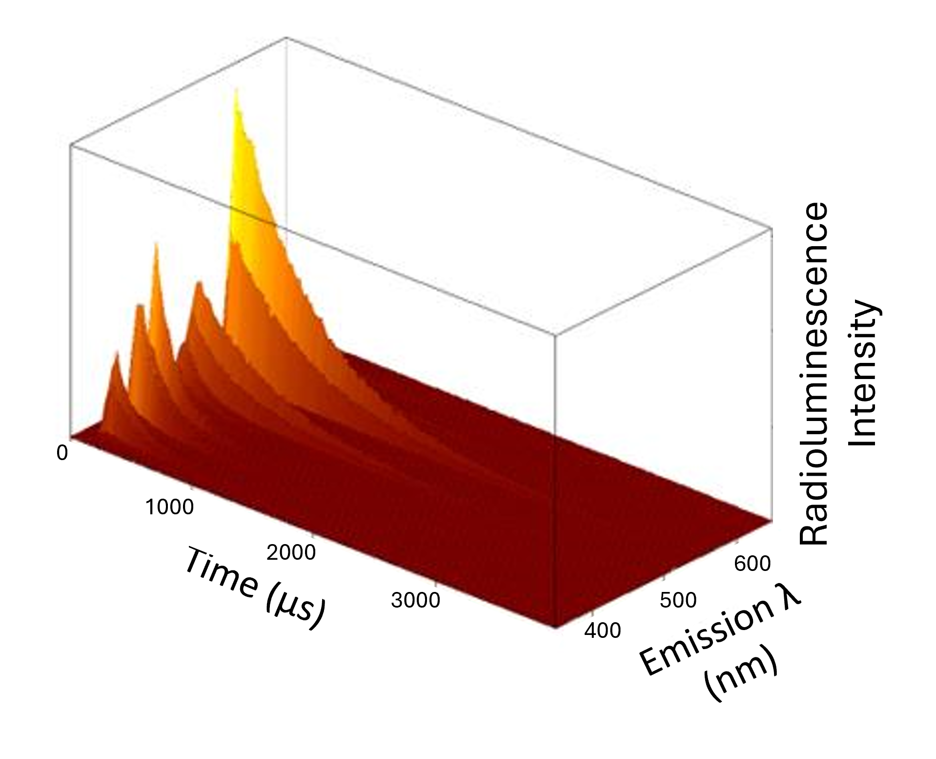
Image Credit: Edinburgh Instruments
How can radioluminescence optimize scintillator materials?
Maria Tesa: Radioluminescence spectroscopy provides key performance metrics such as emission spectra, light yield, linearity, and limit of detection. It also helps identify defects and dopants within the material.
Time-resolved measurements provide insight into decay times, charge carrier dynamics, and material stability. By combining these techniques, you can better understand the photophysics of the scintillator and optimize its composition and structure for improved performance.
What are the practical limits of radioluminescence measurements in terms of time resolution and temperature?
Maria Tesa: The shortest radioluminescence lifetimes you can measure depend on the excitation source and detector. Using a TCSPC setup with a suitable pulsed source, you can typically measure lifetimes down to tens of picoseconds, with excitation pulses as short as around 50 picoseconds.
In terms of temperature, the system can be equipped with a temperature-controlled stage, allowing measurements down to 77 Kelvin and above room temperature. This enables detailed studies of temperature-dependent behavior, which is important for understanding scintillator mechanisms and improving performance.
About Maria Tesa 
Maria Tesa holds a PhD in Physical Chemistry from Heriot-Watt University. She is a Product Manager at Edinburgh Instruments, where she oversees the development and positioning of advanced spectroscopy solutions for research and industrial applications.

This information has been sourced, reviewed, and adapted from materials provided by Edinburgh Instruments.
For more information on this source, please visit Edinburgh Instruments.
Disclaimer: The views expressed here are those of the interviewee and do not necessarily represent the views of AZoM.com Limited (T/A) AZoNetwork, the owner and operator of this website. This disclaimer forms part of the Terms and Conditions of use of this website.