In this interview, industry expert Iona Hill explores how Raman microscopy reveals chemical and structural changes in batteries, enabling real-time insight into degradation, electrolyte behaviour, and failure mechanisms to improve performance and design.
Please can you begin by explaining why Raman microscopy is such a valuable tool for studying electrochemical systems such as batteries?
Raman microscopy is a highly versatile technique because it enables the analysis of a wide range of materials relevant to battery systems, including metal oxides, carbon materials, aqueous electrolytes, and organic electrolytes or gels.
This allows researchers to study all key battery components, from electrodes to electrolytes, as well as processes such as ion transport.
When analyzing electrodes, Raman microscopy can be used to investigate the formation of new phases, structural changes, and degradation products. In electrolytes, it helps examine ion solvation, interfacial interactions, and hydration levels. The technique also enables monitoring of ion insertion and extraction, strain within electrode materials, and the reversibility of electrochemical processes.
Overall, Raman microscopy provides both chemical and structural insights, which are essential for understanding battery performance.
Can you outline how Raman spectroelectrochemistry works in practice?
Raman spectroelectrochemistry combines a Raman microscope with an electrochemical setup, typically consisting of a three-electrode cell and a potentiostat.
The electrochemical cell includes a working electrode, a reference electrode, and a counter electrode, while the potentiostat controls the applied potential and maintains stable electrochemical conditions.
In practice, the electrochemical cell is placed under the Raman microscope, allowing the laser to be focused onto the sample through a window in the cell. While a potential is applied, Raman spectra can be collected simultaneously.
This enables real-time observation of vibrational changes within the material, providing both spatial and temporal insight into electrochemical transformations as they occur.
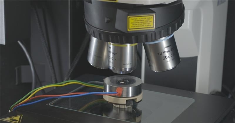
Image Credit: Edinburgh Instruments
What kind of information can a Raman spectrum provide in these studies?
A Raman spectrum provides detailed chemical and structural information that is specific to the material being analysed. The position of Raman bands tells you about chemical structure, while changes in intensity can be linked to concentration variations. The technique can also detect the formation of new phases, impurities, or degradation products.
Additionally, shifts in band position can indicate strain within a material, and changes in band width can reveal information about crystallinity, dopants, or solvation structures. Raman can also be extended to imaging, where spectra are collected across a grid to map spatial variations within a sample, either in situ or ex situ.
What are the key considerations when configuring a Raman microscope for electrochemical measurements?
There are several components to consider, but the most important are the laser, the diffraction grating, and the detector. The laser wavelength must be chosen carefully to balance signal intensity, fluorescence, and resolution.
Shorter wavelengths generally give stronger signals but may introduce fluorescence, whereas near-infrared lasers can minimise fluorescence.
The grating determines spectral resolution, with higher groove densities offering better resolution but a narrower spectral range. Finally, the detector plays a major role in sensitivity.
For example, back-illuminated CCD detectors provide much higher quantum efficiency compared to front-illuminated ones, which is particularly important for low-signal or mapping experiments.
How does detector choice impact Raman imaging performance?
Detector choice has a significant impact on both sensitivity and acquisition speed. Back-illuminated CCD detectors can reach quantum efficiencies of around 95 %, compared to roughly 55 % for front-illuminated detectors. This increased sensitivity allows me to collect stronger signals in shorter acquisition times.
In practical terms, mapping times can be reduced substantially while maintaining data quality. For example, imaging that might take nearly two hours with a standard detector can be reduced to under an hour with a more sensitive one.
Additionally, EMCCD detectors can further enhance signal through electron multiplication, making them ideal for fast or low-light measurements.
Can you give an example of how operando Raman microscopy is applied in electrochemical research?
Operando Raman microscopy can be used to investigate electrocatalysts during reactions such as water splitting. Spectra collected at different applied potentials make it possible for researchers to follow chemical changes in real time.
For example, it can be used to track changes in oxidation states or identify when a material becomes unstable at higher voltages.
This type of analysis helps explain differences in catalytic performance, such as why a material may perform well for hydrogen evolution but not for oxygen evolution. It provides direct insight into how materials behave under real operating conditions.
How can Raman imaging be used to understand electrode degradation?
Raman imaging, especially when combined with chemometric analysis, is a powerful tool for studying structural changes in electrodes before and after cycling. Comparing spatial maps over time reveals how different material phases evolve across the electrode surface.
In composite electrodes, for instance, Raman imaging can highlight the selective degradation of specific phases and show which components remain more stable during repeated cycling. It can also detect deposits such as electrolyte salts forming on the electrode surface.
Compact, research-grade systems like the RM5 are particularly well-suited to this work, offering the spatial resolution and mapping capabilities needed for this type of analysis.
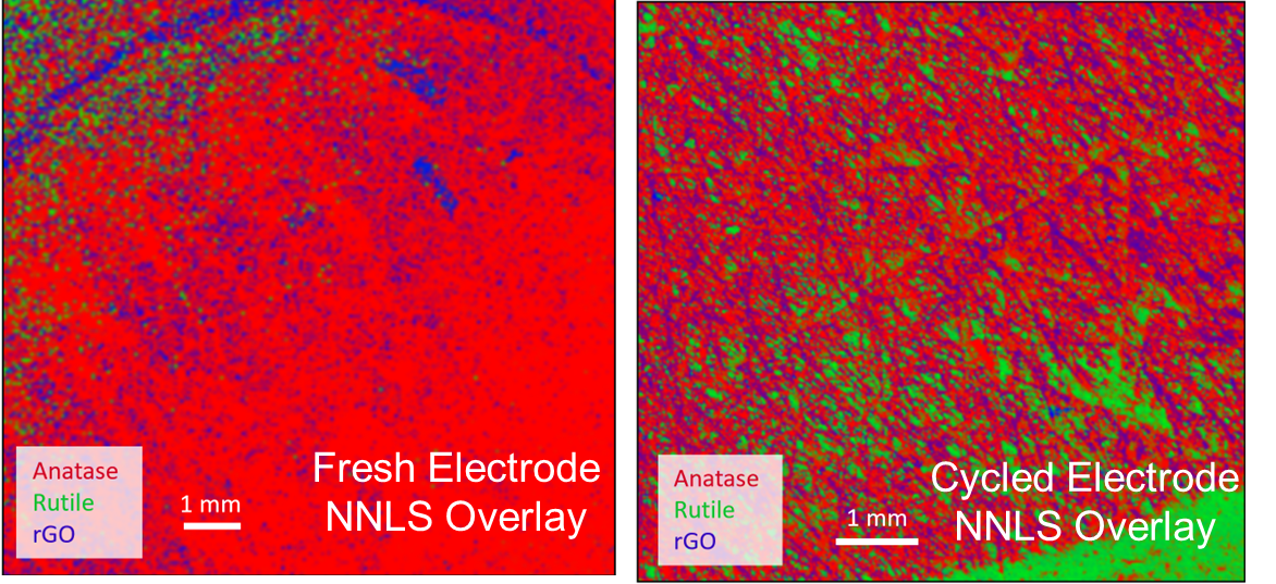
Image Credit: Edinburgh Instruments
How can Raman spectroscopy be used to study electrolyte performance?
Raman spectroscopy can be used to characterise electrolyte structure and understand how modifications affect performance. Analysis of both the fingerprint region and higher wavenumber regions can reveal changes in molecular structure and intermolecular interactions.
One example is hydrogen bonding, which can be monitored through shifts in OH stretching bands. This allows researches to understand how additives influence properties such as flexibility or resistance to freezing.
These insights are particularly important for developing electrolytes that perform well under extreme conditions, such as low temperatures.
Can Raman spectroscopy be used to monitor solid electrolyte interface (SEI) formation?
Yes, Raman spectroscopy can absolutely be used to monitor the formation of the solid electrolyte interface. In systems like lithium batteries, the SEI often contains species such as lithium carbonate or lithium fluoride, both of which have distinct vibrational signatures.
Operando Raman measurements allow the appearence and evolution of these species to be tracked during battery cycling which makes it possible to observe SEI formation and growth directly.
Bringing it all together, how can Raman spectroscopy help diagnose battery failure?
Raman spectroscopy is very useful for diagnosing battery failure as it can analyze multiple components of the system, including the electrode, electrolyte, and interfaces. This is key becasue in many cases, failures are caused by a combination of factors, such as structural degradation, phase changes, or unwanted side reactions.
By examining spectral changes across these components, researchers can identify degradation pathways, detect new or unstable phases, and observe electrolyte decomposition or deposition. This holistic view makes it possible to understand why a battery is losing capacity and how its design can be improved.
About Iona Hill 
Iona Hill holds a PhD in Chemistry from the University of Strathclyde, following an MChem in Forensic and Analytical Chemistry. She is currently an Application Scientist at Edinburgh Instruments, specialising in Raman spectroscopy, instrument optimisation, and advanced analytical support for research and industrial applications.

This information has been sourced, reviewed, and adapted from materials provided by Edinburgh Instruments.
For more information on this source, please visit Edinburgh Instruments.
Disclaimer: The views expressed here are those of the interviewee and do not necessarily represent the views of AZoM.com Limited (T/A) AZoNetwork, the owner and operator of this website. This disclaimer forms part of the Terms and Conditions of use of this website.