From SemplorReviewed by Olivia FrostSep 30 2026
This article is based on a poster originally authored by Dr. Ivan Lobato, Eric Toering, Dr. Serge Gavras, and Prof. Dr. Karel van der Mast from Semplor.
EDS spectra comprise characteristic X-ray peaks superimposed on a Bremsstrahlung continuum. The peaks encode elemental composition, whereas the continuum contains complementary information associated with the local mean-Z and specimen geometry. At low counts, signal-dependent Poisson noise obscures weak peaks and complicates quantitative spectral reconstruction.
Detector physics introduces additional structured ambiguities. Si-escape peaks occur 1.74 keV below energetic parent lines, while pulse pile-up at high count rates produces sum peaks at E1 +E2. Sequential denoising and background subtraction neither preserve the separated peak and continuum components nor explicitly resolve them at the detector stage.
Objective: Jointly denoise EDS spectra, separate characteristic peaks from the Bremsstrahlung background, and compensate for Si escape and pulse pile-up in a single physics-structured inverse problem
Method: Physics-Structured Targets and One Coupled Network
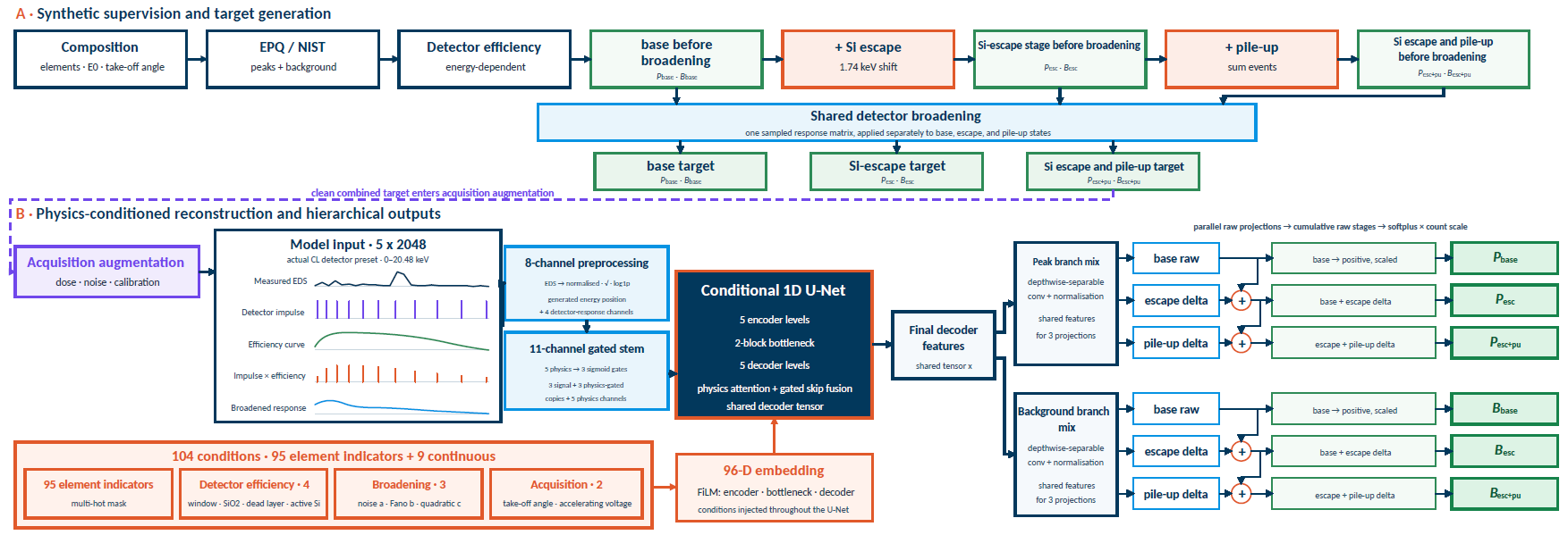
Image Credit: Semplor
Stage notation: P and B denote the peak and Bremsstrahlung components, respectively. Pbase/Bbase: Si escape and pile-up removed; Pesc/Besc: Si escape retained and pile-up removed; Pesc+pu/Besc+pu: both artifacts retained before stochastic acquisition augmentation.
Results 1: Fe2O3 Spectra Validate Low-Dose Reconstruction and Detector-Stage Separation
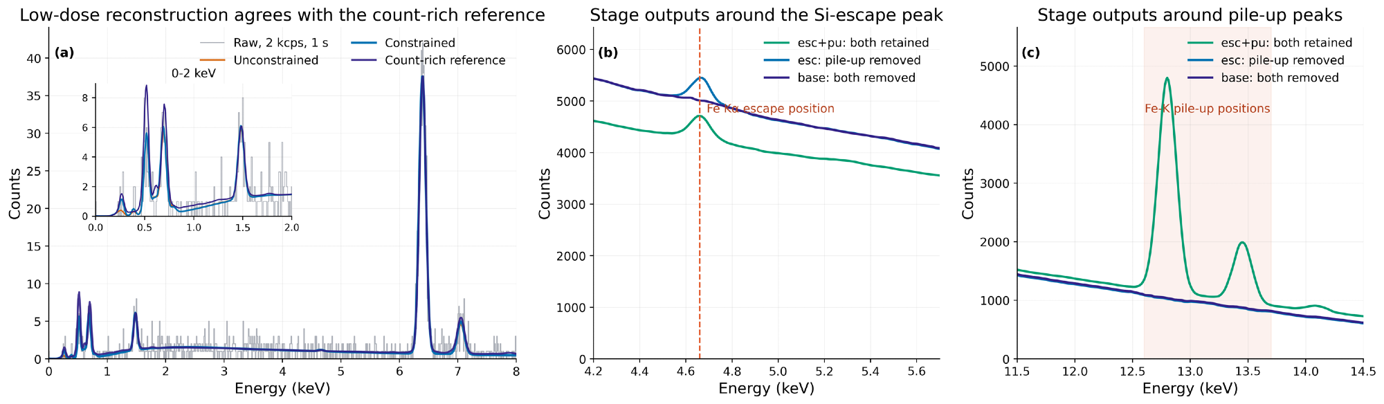
Image Credit: Semplor
At 20 kV, the constrained 2 kcps, 1 s reconstruction agrees with a count-rich reference. The stage-resolved outputs identify the Fe Kα Si-escape position and the Fe K pile-up region.
Results 2: Signal Separation Across the Thermite Spectral Image
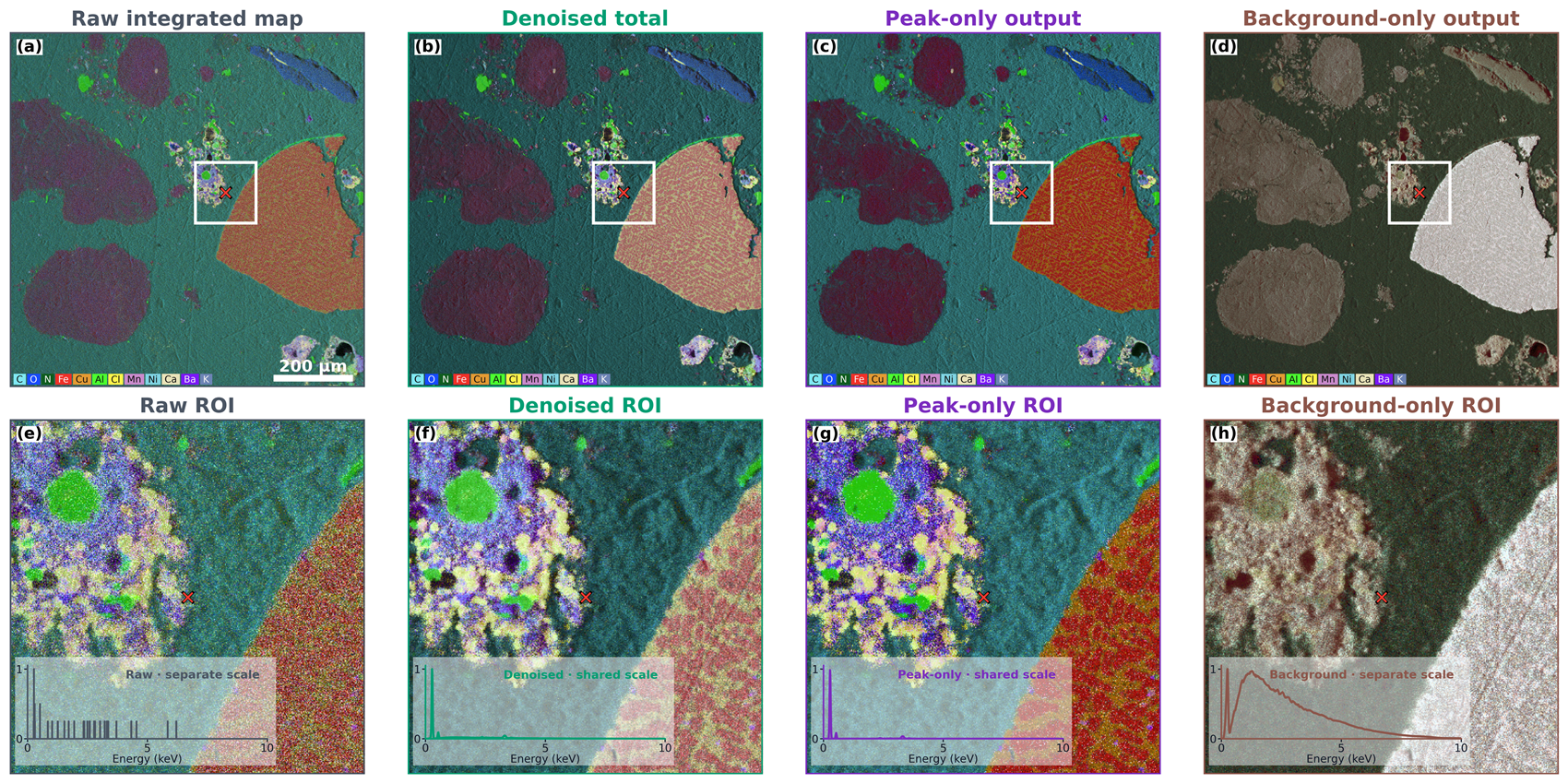
Image Credit: Semplor
Each column links one full-field map to its marked ROI and central-pixel spectrum. Denoised and peak-only insets share the denoised-total scale; raw counts and background are normalized separately so their spectral shapes remain visible.
Conclusions
- Physics-structured targets, staged losses, and one shared conditional network separate low-dose denoising from detector-artifact compensation; the method is not specific to a single acquisition condition.
- Six coupled outputs from two hierarchical branches recover matched peak/background pairs at three cumulative physical stages: both Si escape and pile-up retained, Si escape retained with pile-up removed, and both artifacts removed.
- The Fe2O3 experiments span complementary count regimes: conditioning stabilizes low-dose reconstruction; constrained and unconstrained solutions converge as counts increase; and differences between stages reveal Si-escape and pile-up structure at high count rates.
- The thermite spectral image shows that denoising suppresses speckle, peak-only reconstruction strengthens compositional contrast, and the separated background retains complementary mean-Z and morphological contrast. The model supports constrained, semi-constrained, and unconditional inference.
References and Further Reading
- Pouchou, J.-L. and Pichoir, F. (1991). Quantitative Analysis of Homogeneous or Stratified Microvolumes Applying the Model “PAP.” Electron Probe Quantitation, pp.31–75. DOI:10.1007/978-1-4899-2617-3_4. https://link.springer.com/chapter/10.1007/978-1-4899-2617-3_4.
- Ritchie, N.W.M. (2009). Spectrum Simulation in DTSA-II. Microscopy and Microanalysis, 15(5), pp.454–468. DOI:10.1017/s1431927609990407. https://academic.oup.com/mam/article-abstract/15/5/454/6919550.
- Schoonjans, T., et al. (2011). The xraylib library for X-ray–matter interactions. Recent developments. Spectrochimica Acta Part B: Atomic Spectroscopy, 66(11), pp.776–784. DOI:10.1016/j.sab.2011.09.011. https://www.sciencedirect.com/science/article/abs/pii/S0584854711001984.
- Ronneberger, O., Fischer, P. and Brox, T. (2015). U-Net: Convolutional Networks for Biomedical Image Segmentation. Lecture Notes in Computer Science, 9351, pp.234–241. DOI:10.1007/978-3-319-24574-4_28. https://link.springer.com/chapter/10.1007/978-3-319-24574-4_28.
- Semplor BV. NANOS tabletop SEM product information (2026).

This information has been sourced, reviewed, and adapted from materials provided by Semplor.
For more information on this source, please visit Semplor.