Please can you introduce yourself, tell us a little about your role at XEI Scientific, and explain how your work relates to contamination control in microscopy?
I’m Michal, and I’ve worked in electron microscopy for almost 30 years - ever since completing my doctorate at the University of Tokyo. I began my professional career as a service engineer at Gatan in Japan and later led its business in Asia. After spending three years as the president of TESCAN USA, I returned to Gatan and EDAX as the global vice-president of sales.
When the opportunity arose to acquire XEI Scientific, I partnered with my colleague and friend, Dr. Christopher Booth, to continue building on the company's long history of innovation in contamination control.
At XEI Scientific, we focus on two simple but important questions: how do we help researchers obtain reliable, reproducible data from their microscopes? How do we aid their productivity?
Contamination control is a critical part of that answer. Even the most advanced microscope cannot compensate for an unwanted contamination layer on the specimen surface. If the sample or the vacuum environment is contaminated, image quality, analytical accuracy, and the confidence in the results will all suffer.
To set the scene, what exactly is hydrocarbon contamination in microscopy, and where does it typically come from?
Hydrocarbon contamination is usually a two-step process. First, organic molecules accumulate on a specimen's surface. Then, under electron- or ion-beam irradiation, those molecules decompose and polymerize into a stable carbon-rich deposit.
We often use the term "hydrocarbon contamination" as a shorthand, although these residues may contain oxygen and other elements in addition to carbon and hydrogen.
Common sources of contamination include sample-preparation chemicals, adhesives, solvents, handling residues, laboratory air, and outgassing from specimens or microscope components. Historically, vacuum pump oils were also a major contributor, although modern vacuum systems have significantly reduced this source.
The challenge becomes apparent once the beam is introduced. Adsorbed molecules can fragment and crosslink under the irradiation, leaving behind a carbonaceous deposit precisely in the area under examination. Additional molecules may continue arriving from the gas phase or migrate across the surface, allowing the contamination layer to grow during imaging or analysis. At that point, microscopy is no longer simply observing the sample. The act of imaging is actively modifying the surface being studied.
How can researchers tell when hydrocarbon contamination is present, and are there cases where it may be affecting results without being immediately obvious?
One of the most recognizable warning signs is the appearance of a scan footprint. When the magnification is reduced, a previously scanned area may appear as a dark rectangle in an SEM image. The exact contrast depends on the specimen and imaging conditions, but experienced microscopists will immediately recognize this as evidence of beam-induced contamination.
There is a useful practical observation. If the entire scanned region darkens, the contamination often originates from residual hydrocarbons within the microscope chamber. If contamination appears primarily around the perimeter of the scanned area, the specimen itself is likely the source.
The more challenging cases are those that remain invisible. A surface may appear perfectly clean while still carrying enough organic residue to influence analytical measurements. Surface-sensitive techniques such as EDS, EBSD, XPS, Auger spectroscopy, and SIMS can be affected long before contamination becomes obvious in an image. For that reason, a visually clean image should never be assumed to represent a chemically clean surface.
Why is hydrocarbon contamination something microscopy users should be paying closer attention to?
When a laboratory invests in an advanced microscope, it is buying the ability to resolve smaller features, measure more accurately, and answer difficult questions. Hydrocarbon contamination directly undermines the return on investment on that expensive tool.
The critical point is that contamination builds in the very region being measured. As the beam interacts with residual hydrocarbons, it can create a carbonaceous deposit on the specimen - actively altering the surface mid-measurement.
This makes contamination control a core requirement for data quality. A sharp-looking image alone does not establish that contamination is adequately controlled.
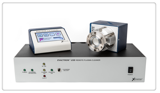
Image Credit: XEI Scientific, Inc.
What impact can contamination have on image quality, analytical results, resolution, and the overall reliability of microscopy data?
Beam scanning causes a dark rectangle to burn over the sample, obscuring features. The contamination buildup creates an effective barrier that broadens the probe profile before it even hits the sample, reducing the effective spatial resolution of both SEM and TEM. Furthermore, hydrocarbon layers are insulating. As the electron beam scans a contaminated area, charge builds up locally, leading to image drift and erratic beam deflections.
Even a thin layer of carbonaceous contamination compromises analytical accuracy. In EDS, it introduces an unwanted carbon signal and attenuates low-energy X-rays from the underlying specimen. In EELS, it adds carbon-related spectral features and increases electron scattering, raising the background and obscuring signals of interest. These effects can bias composition measurements and make weak signals harder to interpret.
Similarly, electron backscatter diffraction (EBSD) relies on electrons escaping from the top few nanometers of the crystal lattice. A carbon film scatters these electrons inelastically, degrading pattern quality, lowering the indexing rate, and preventing accurate orientation mapping or phase identification.
Contamination also impacts vacuum levels and pump-down times, adding constrains to microscope operations and degrading the throughput.
Ultimately, contamination introduces uncertainty into measurements and can reduce confidence in the obtained data.
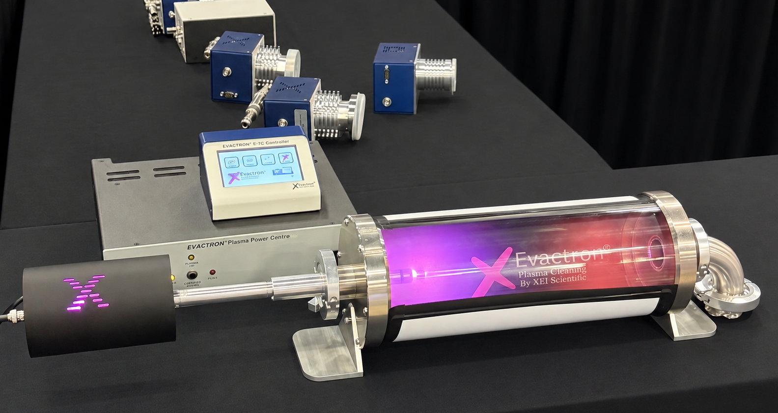 Image Credit: XEI Scientific, Inc.
Can you share some examples of how hydrocarbon contamination has affected particular laboratories, microscopy techniques, or industrial applications?
For decades, electron microscopists struggled against the ghost in the machine. Whenever they focused the electron beam to a fine probe or scanned a part of the sample, a dark spot or area would appear. The culprits were the traces of pump oil, fingerprints, and plasticizers floating inside the vacuum chamber.
The electron beam would crack these and polymerize them. One could describe it as unintentionally 3D-printing amorphous carbon with an electron beam. To solve the problem, XEI Scientific founder, Mr. Ronald Vane, introduced the world's first commercial plasma cleaner in 1999.
Historically, many laboratories relied on liquid-nitrogen cold traps to capture contamination before it reached the specimen. Mr. Vane’s hollow cathode design fundamentally changed contamination-control workflows by providing a more direct and efficient way of removing organic residues from the vacuum environment.
When we talk about the “real cost” of contamination, what should we be considering beyond the immediate impact on the image or dataset?
The real cost of contamination extends well beyond a compromised image or dataset. It accumulates through lost instrument time, repeated sample handling, additional measurements, troubleshooting, reduced throughput, and avoidable service calls. Every hour spent repeating work is an hour unavailable for advancing research or supporting another customer.
Consider an illustrative calculation. At an instrument rate of $300 per hour, recovering just two otherwise lost hours each week represents $31,200 in annual instrument capacity. Against a plasma cleaner investment of approximately $30,000, that recovered capacity alone could justify the purchase within about a year. That is why I view effective contamination control as a productivity investment, not maintenance cost.
There is also a human cost that hourly rates do not capture: losing a valuable region of interest, repeating sample preparation or extra time spent on imaging and analysis. Reliable measurements protect researchers and laboratories give confidence in the data and protect their professional reputation.
What methods are currently available to reduce or eliminate hydrocarbon contamination, and what are the strengths and limitations of each approach?
Prevention remains the first line of defense. Good sample-handling practices, clean tools, cleanroom gloves, and minimizing unnecessary exposure to airborne contaminants can all reduce contamination levels before a specimen enters the microscope. Nevertheless, even well-controlled laboratories accumulate trace levels of organic contamination over time, making active cleaning an essential part of microscopy workflows.
One common way to clean a vacuum chamber is using ozone or UV treatment. While ozone processing is often mistakenly called plasma cleaning, the fundamental chemistries and effectiveness of the two differ significantly.
In-situ plasma systems generate a high-density flux of oxygen radicals. These are highly reactive species with unpaired electrons capable of rapidly cleaving C–H and C–C bonds to transform contaminants into volatile, easily pumped-out species. In contrast, molecular ozone is far less reactive on its own. It requires thermal or photon-assisted decomposition to yield reactive oxygen, rendering ozone significantly less efficient.
The mechanism of radical generation also plays a pivotal role in process control. Capacitively coupled plasma (CCP) generates an abundance of reactive gas radicals that chemically dissociate organic residues. Because CCP operates as a cold plasma (around 50 °C) , it prevents sample thermal damage. Additionally, hollow cathode design reduces energetic ion bombardment, minimizing unwanted physical etching of sensitive samples and chamber components.
Users understandably ask what the best cleaning recipe is. We found that breaking it up into shorter intervals (e.g., three sets of five minutes with a one-minute pause) is more effective than continuous plasma exposure for the same total duration. This is not about damaging components but about removing volatile molecules that may have reattached themselves to the vacuum chamber during cleaning.
Are there particular samples, techniques, or microscopy environments where contamination is particularly important to manage? What are the challenges?
Contamination control is particularly important when the contamination layer is significant relative to the feature, specimen thickness, or signal being measured. That includes (S)TEM, high-resolution SEM, and surface-sensitive techniques such as Auger spectroscopy, XPS, and SIMS. Ultrathin specimens, nanoparticles, and two-dimensional materials are especially challenging because even a small amount of contamination can obscure the structure or chemistry of interest.
Take transmission electron microscopes for illustration. Keeping the TEMs free of contamination remains a persistent challenge. Users commonly attempt a "beam shower," flooding a large area with a high-current electron beam. This treatment merely polymerizes hydrocarbons fixing them in place rather than removing it. Cold traps require refilling liquid nitrogen and thermal cycling. Again, the contamination itself remains inside the column. Column bakeouts can sideline an instrument for days and require manufacturer's involvement. XEI Scientific's TEM Wand™ offers a fundamentally different solution: a side-entry holder that allows users to clean the interior of their (S)TEM by the user - directly, quickly, and effectively, without lengthy downtime or service calls.
Looking to the future, what advances in materials, instrument design, cleaning technologies, or contamination monitoring could help reduce the impact of hydrocarbon contamination in microscopy?
The future may bring shift from scheduled cleaning to evidence-based contamination control: measuring contamination, cleaning where needed, and verifying the result. Residual-gas analysis, controlled deposition tests, and closer integration with microscope controls could make this a routine part of the workflow.
Selectivity remains essential, particularly for sensitive materials. Location targeted delivery of reactive species, alternative gas chemistries, and carefully controlled power and exposure times offer ways to remove unwanted residues while preserving the material under study.
Ultimately, the goal is not to maximize cleaning intensity. Rather, it is to achieve the highest possible measurement reliability with the smallest possible impact on the material being studied. The contamination control should become as fundamental as instrument alignment and calibration.
About Michal Rabara 
Michal's career began with superconducting materials at the University of Tokyo and grew through hands-on work helping scientists get the most from their electron microscopes. Starting as a service engineer in Asia, he later managed regional organizations before becoming global sales VP at Gatan and EDAX and serving as the President of Tescan USA. Living and working in Europe, in Japan, Singapore, and the United States - and speaking multiple languages - has given him a broad perspective on connecting with people across different cultures. Today, as owner and Chief Commercial Officer of XEI Scientific, Michal brings a researcher’s curiosity and almost three decades of experience to helping scientists explore materials and technologies.
Driven by a passion for innovation and advancing microscopy technology, Michal partnered with long-time colleague Dr. Christopher Booth to acquire Evactron building on its pioneering legacy in contamination control and shaping its next chapter of growth.
This information has been sourced, reviewed, and adapted from materials provided by XEI Scientific, Inc.
For more information on this source, please visit XEI Scientific, Inc.
Disclaimer: The views expressed here are those of the interviewee and do not necessarily represent the views of AZoM.com Limited (T/A) AZoNetwork, the owner and operator of this website. This disclaimer forms part of the Terms and Conditions of use of this website.