The column design of scanning electron microscopes (SEMs) has included an electron gun, objective lens, an aperture or aperture strip, and a condenser lens or two. An additional lens incorporated into the column on top of the objective lens by TESCAN enables new imaging modes and improved performance to LaB6, tungsten-source, and field emission SEMs. This additional lens is termed as intermediate lens (IML).
IML Modes
Different IML modes available are listed as below:
- Resolution mode for high-resolution imaging
- Field mode for large field of view imaging
- Depth mode for high depth of focus imaging
- Beam Rocking mode for Selected Area Electron Channeling and live 3D stereoscopic imaging (3D mode)
- Wide Field mode for wide field of view imaging and ultra-low magnification
A click of the mouse will allow switching between the aforementioned imaging modes as they are fully automated and computer controlled modes. As a result, components in the column can be selected, installed, or mechanically aligned without any operator interference. In addition, the sample working distance can be changed automatically. Figure 1 shows the column design for each imaging mode.
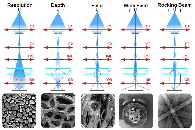
Figure 1. Different IML modes
Field Mode
The magnification range of TESCAN SEMs is the broadest available on a commercial SEM, from 1x to 1,000,000x. This means that both ultra-low magnification “macro” imaging and high-resolution imaging are possible with TESCAN SEMs. The SEM is capable of scanning a field of view up to 15mm at the analytical working distance and beyond 100mm at long working distances.
Depth Mode
With TESCAN’s IML design, the microscope can reach a high depth of focus of more than 100mm, a value far beyond the working distance range of the stage. This is suitable for analysis of samples such as foams having cells in multiple layers and rough samples.
Wide Field Mode
The SEM is said to be in wide field mode because the IML acts as the objective lens when the objective lens is turned off. Since the IML is placed above the objective lens in the column, it is farther away from the sample surface when compared to the objective lens. As a result, IML yields magnifications down to 1x by focusing over a larger area.
Rocking Beam Mode for Electron Channeling
In the Beam Rocking Mode, the electron beam can be rocked at different angles to a spot on the sample. This action will produce a Kikuchi pattern as shown in Figure 2. These patterns are connected with the crystalline planes in samples being analyzed, revealing their composition and cell structure.
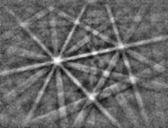
Figure 2. Kikuchi patterns
Beam Rocking Mode for Live 3D Imaging
The TESCAN SEM is the only commercially available SEM that can capture and archive live 3D stereoscopic images (Figure 3). Live 3D images can be viewed while changing magnifications and navigating around the sample. Moreover, the TESCAN SEM is the only SEM that can provide integrated quantitative 3D reconstruction and analysis.
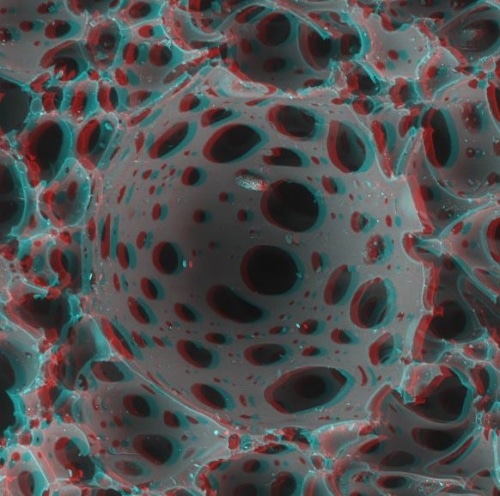
Figure 3. An example for live 3D stereoscopic imaging
Quantitative digital elevation maps can be created from stereo image pairs, thanks to the fully integrated software functionality provided by the optional integrated MeXpackage. The TESCAN SEM acquires stereo images automatically using beam tilting technology.
Comprehensive 3D renderings of the sample surface, shaded with the actual SEM image, can be created with the system. In addition, the 3D model can be zoomed, panned, tilted and rotated. Furthermore, quantitative volume, area, and profile measurements can be extracted from the digital elevation maps.
TESCAN Group
Founded in 1991 by a group of managers and engineers from Tesla with its electron microscopy history starting in the 1950’s, today TESCAN is a globally renowned supplier of Focused Ion Beam workstations, Scanning Electron Microscopes and Optical Microscopes. TESCAN’s innovative solutions and collaborative nature with its customers have won it a leading position in the world of nano- and microtechnology. The company is proud to participate in premier research projects with prominent institutions across a range of scientific fields. TESCAN provides its clients with leading-class products in terms of value, quality and reliability. TESCAN Group is the North American arm of TESCAN Group, a multinational company established by the merger of Czech company TESCAN, a leading global supplier of SEMs and Focused Ion Beam workstations, and the French company ORSAY PHYSICS, a world leader in customized Focused Ion Beam and Electron Beam technology.

This information has been sourced, reviewed and adapted from materials provided by TESCAN Group.
For more information on this source, please visit TESCAN Group.