Over 50 years ago, the first commercially available SEM was unveiled. Yet, to this day no internationally accepted standard exists for the determination of SEM resolution. Further, to confuse matters even more, each SEM manufacturer depends on their own sample and methods for establishing resolution.
Two of the most popular ways to determine resolution are:
- Measuring the separation between two adjacent objects
- Acquisition of a line profile of the signal intensity changes across a sharp edge
Traditionally, JEOL has applied a sample of small gold particles evaporated on a carbon substrate (Figure 1). With this kind of sample, resolution is then established via calculating the separation between two adjacent gold particles, or by examining the line profile of the changes in signal intensity across the edge of a gold particle.
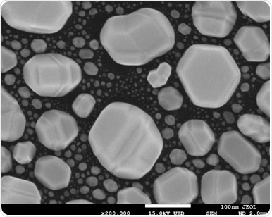
Figure 1. SEM Image of a gold on carbon test sample. Image Credit: JEOL USA, Inc.
Each of these methods is subjective in nature. Determining what constitutes as the particle’s edge will differ from person to person based on individual interpretation or how each person comes to observe the edge of a particle. When conducting the line profile technique, the distance for the signal transition is deemed to be related to probe diameter.
JEOL uses the conventional protocol and measures at the 84th and 16th percentile of the transition (1 sigma value). This is not true for all manufacturers and values of 75th/25th and 65th/35th percentile have been declared by other vendors. As illustrated in Table 1, this causes lower reported values of resolution even from the same edge profile in an image. Evidently, it is not easy to compare resolution specifications today between different manufacturers.
Table 1. Example showing the resolution determined from the same edge profile using three different percentiles to calculate the beam diameter. Source: JEOL USA, Inc.
| |
84% |
75% |
65% |
| Beam diameter (resolution) |
1.4 nm |
1.1 nm |
0.6 nm |
An additional layer of complexity comes with digital image acquisition in contemporary SEM microscopes. The final image pixel resolution influences the smallest features that can be resolved, this means that the resolution measurement is closely linked to the pixel size. Regarding an image captured at 100,000X with a field of view of 1.28 um: if the resolution of the image pixel is 1280 X 960, then a pixel length of 1 nm/pixel is taken. To identify a probe diameter of 3 nm would necessitate image information that could be seen over just 3 pixels. Yet, capturing the same image with an enhanced pixel density of 2560 x 1920 would mean that the equivalent information could be seen across 6 pixels.
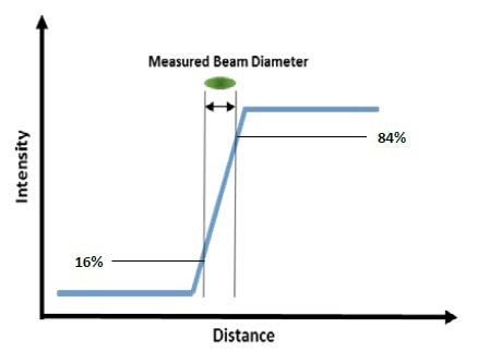
Figure 2. Line Profile Method for SEM Resolution. Image Credit: JEOL USA, Inc.
Due to the fact image acquisition and the effective measurement procedures can differ substantially between various electron microscope vendors, it is crucial to bear this in mind when relating and comparing resolution numbers. How the measurement is made and what sample and parameters are selected can have a profound impact on the resolution number reported.

This information has been sourced, reviewed and adapted from materials provided by JEOL USA, Inc.
For more information on this source, please visit JEOL USA, Inc.