Visualization of the nano-world is a key component in many modern academic institutions. However, to harness the power of this expansive technique, as well as other essentials of cutting-edge research, a core facility where instrumentation and researchers come together to serve the advancement of science is crucial.
To conceptualize and build such a core facility is certainly not an easy task, although some make the creation of this synergistic environment feel like a walk in the park, such as the George Washington University.
In 2015, The George Washington University (GW) had a vision for a brand-new Science and Engineering Hall that would co-locate various shared facilities and disciplines of scientific research into a single, integrated collaborative environment.
A core facility for materials characterization and nanofabrication was a key component. The goal of this facility was to serve researchers in Chemistry, Physics, Biology, and Anthropology as well as GW’s School of Engineering and Applied Science, School of Medicine and Health Sciences, and the Milken Institute School of Public Health.
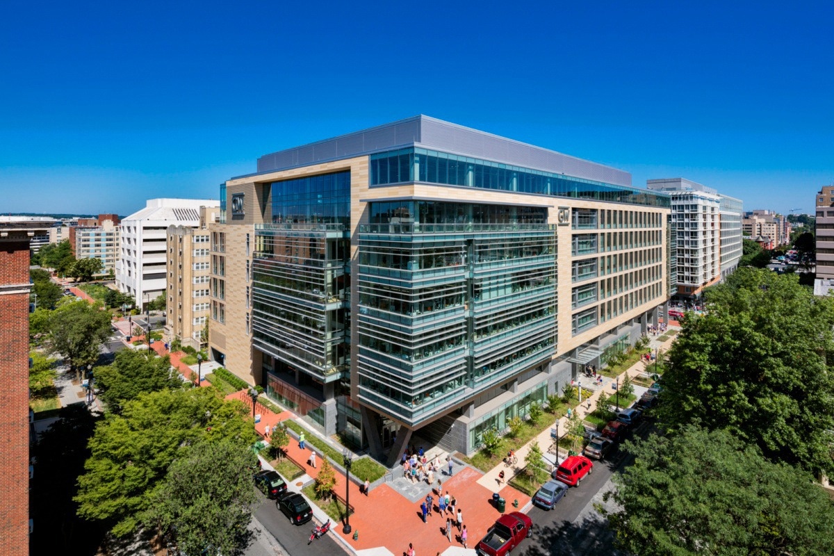
George Washington University – Science and Engineering Hall. Image Credit: Albert Vecerka
What were the main objectives behind establishing a core facility featuring electron microscopy on the GW campus?
With a new building, Dr. Anastas Popratiloff, Director and Lead Scientist of the GW Nanofabrication and Imaging Center saw an opportunity to bring modern electron microscopy (EM) to GW’s campus and empower users with the cutting-edge, novel research techniques made possible through imaging and analysis.
One aspect of EM that was particularly intriguing for GW was that of automation and correlative workflows, along with the ability to seamlessly utilize multiple modalities on a single platform. The goal was to make electron microscopy more desirable and approachable for users from all disciplines.
What equipment is available in GW’s state-of-the-art facility and how does this benefit the scientific community?
Flexibility for multimodal imaging and analysis within as well as across platforms is the key to modern EM-related science. GW’s impressive lineup of a Thermo Scientific Teneo scanning electron microscope (SEM), Helios focued ion beam scanning electron microscope (FIB-SEM), Talos F200X transmission electron microscope (TEM), and Maps correlative workflow software suite, along with many state-of-the-art optical microscopes and even a cleanroom, enables researchers from the university as well as surrounding institutions to shed new light (as well as electrons) on challenging questions.
"Combining light and electron microscopy is a differentiator. Most improvements in life science were achieved because we used materials science techniques." said Dr. Popratiloff.
Thermo Scientific Maps software unlocked such approaches by allowing users to capture low-magnification navigation images from one instrument, then stitch high-resolution images from another to see the entire picture (i.e., global context and resolution).
In speaking about Maps, Dr. Popratiloff went on to say, "This was a game-changer. You don’t just get a single image and walk away. Now, scientists from across the university are coming here to do electron microscopy, as well as users [in the National Capital Region] and beyond."
By pushing the envelope in using Maps and scanning transmission electron microscopy (STEM), GW has been able to image as well as quantify exosomes and other vesicles effectively.
In one case, 1500 images were acquired on the Talos and stitched together into one large image, allowing analysis on a much larger and more definitive scale than others.
The Talos is ridiculously stable at 200kV... no drift, nothing.
Dr. Anastas Popratiloff, Director and Lead Scientist of the GW Nanofabrication and Imaging Center
During a recent visit from Product Applications Specialist Alex Hall of Thermo Fisher, the team at GW also experienced aspects of the 3D Wizard, deep learning and prediction, multichannel data handling, segmentation, correlative light and electron microscopy (CLEM), and other techniques so that they can leverage these to the fullest extent.
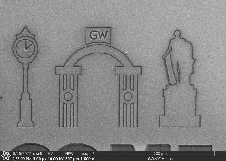
FIB-SEM art using the Thermo Scientific Helios system. Image Credit: George Washington University Nanofabrication and Imaging Center
How did the virtual environment and COVID-19 pandemic affect GW’s instrumentation operations?
Being able to run the microscopes remotely is standard for Thermo Scientific microscopes. However, the full extent of the importance of these reliable capabilities were not fully realized until the pandemic hit in 2020.
"When COVID hit, the university shifted activities to a virtual modality, but we were able to keep working. I was running the whole facility [remotely]. This is how I envisioned things would work", said Dr. Popratiloff.
The university was able to run tests that lasted 16 hours+ without an issue, and, in one instance, they ran tests for two and a half days. "The microscope just kept capturing and stitching, and capturing", Dr. Popratiloff said. He also went on to collect preliminary data for a grant with a pressing deadline, all while much of the world was at a standstill.
It did not stop there. Collaboration continued in the virtual space, as it was very important to make scientific progress even though many interactions could not take place in person. GW has also worked with investigators from George Mason University and others using this remote environment.
What were some of the challenges involved in establishing a successful core facility?
Making the facility at GW what it is today required a lot of high-level leadership, cross-unit collaboration, and a significant amount of institutional investment. Instrumentation grants are more challenging and competitive these days. A word of advice for those looking to establish similar programs is to create a hub for intellectual and experimental expansion.
Data is crucial, and one must justify as well as interlink all the pieces of the puzzle.
What is on the horizon for GW?
GW will be hosting its annual CLEM workshop this year, from April 24th-28th, 2023.
"We call it correlative, but it is actually multimodal microscopy and we train the next generation of technically savvy microscopists to create knowledge transfer as they further science", said Dr. Popratiloff. He is also looking into a Thermo Scientific VolumeScope + FIB-SEM + 3D EDS approach as these systems can be used to address some unique questions and synergistic science where a functional community is built in order to help others grow.
Check out the upcoming workshop HERE.
About Dr. Anastas Popratiloff 
Anastas Popratiloff, MD, PhD is the Director and Lead Research Scientist for the GW Nanofabrication and Imaging Center (GWNIC) and limited service professor of Anatomy and Cell Biology at GW School of Medicine & Health Sciences. His current research focuses on neuronal structure, neuronal networks, and models of major neurological conditions. He received his MD degree from the Medical University of Sofia in Bulgaria and his doctorate from the Faculty of Medicine at the University of Cologne in Germany. From 1994-1997 he received postdoctoral training at the laboratory of Prof. Aldo Rustioni. It was here that he conducted pioneering research on the role of AMPA receptors at the first brain synapses processing pain and AMPA homeostatic regulation in sensory deprivation. Prior to joining GW, he held academic research and teaching positions at the Medical University of Sofia and the University of Cologne. He was appointed the Director of the GW Center for Microscopy and Image Analysis and Research Associate Professor of Anatomy in 2006, and served as co-director of the Cell and Tissue Imaging Core of the District of Columbia Intellectual and Developmental Disabilities Research Center. He took a leading role in the design and implementation of the GWNIC in 2014.
This information has been sourced, reviewed and adapted from materials provided by Thermo Fisher Scientific – Electron Microscopy Solutions.
For more information on this source, please visit Thermo Fisher Scientific – Electron Microscopy Solutions.
Disclaimer: The views expressed here are those of the interviewee and do not necessarily represent the views of AZoM.com Limited (T/A) AZoNetwork, the owner and operator of this website. This disclaimer forms part of the Terms and Conditions of use of this website.