In this interview, AZoM talks to Andrew Schmitz of Thermo Fisher Scientific, about the analysis of micro layers through FTIR microscopy and its impact on the scientific industry.
Please can you introduce yourself and FTIR microscopy?
My name is Andrew Schmitz and I am going to introduce the analysis of micro layers via FTIR microscopy using a Nicolet RaptIR FTIR Microscope.
In micro-level FTIR microscopy, samples can be less than a hundred microns, such as some microplastics, inclusions, or contaminants embedded into bulk materials.
In microscopy, you have three main techniques that are similar to macro-FTIR, including reflection, attenuated total reflectance (ATR), and transmission. Reflection is the infrared energy reflecting off a sample. With transmission, the infrared energy is transmitted through the sample. ATR uses the micro-ATR tip. As with micro-FTIR, this can be non-destructive.
With microscopy, you need point-by-point analysis or mapping. With point-by-point, you can look at specific samples on the slide or a filter.
With mapping, you can look at a section of a material that consists of multiple chemical components, such as a paint chip or laminate, or a sample in the lab that has a mixture of cellulose and polyethylene. This technique is great for quality control, forensics, or chemical identification.
What are the features of the Nicolet RaptIR FTIR Microscope?
The Thermo Scientific™ Nicolet™ RaptIR™ FTIR Microscope couples with the Thermo Scientific™ Nicolet™ iS50 FTIR Spectrometer and benefits from a 6-megapixel camera.
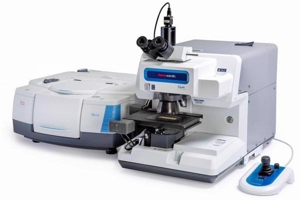
Image credit: Thermo Scientific - Chemical Analysis
It features a motorized nose piece that enables easy change in between objectives as opposed to manually rotating it. There are two objectives on this motor nose piece, a 4x and a 15x. The 4x gives a wide field of view for finding samples easier, and the 15x zooms in on a target.
There are also optional visual polarizers for any polarized measurements.
The Thermo Scientific™ OMNIC™ Paradigm Software powers the microscope. This software is an advanced version of the prior OMNIC software. It provides quicker and easier data collection, with sessions that can collect multiple-point measurements and maps.
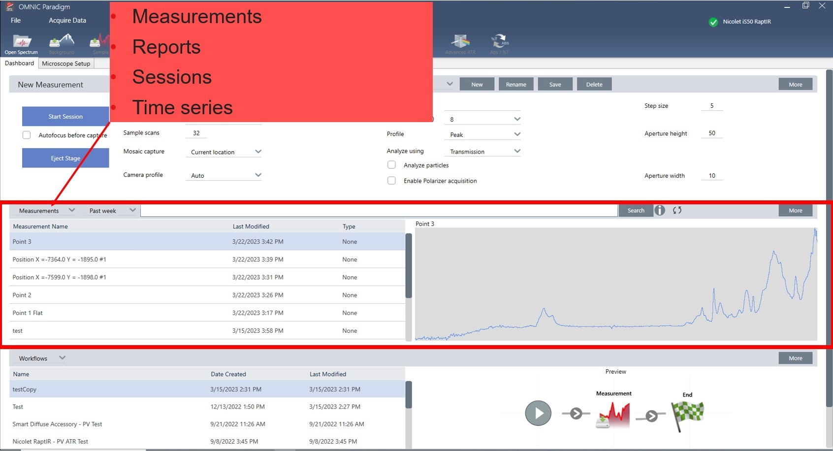
Image credit: Thermo Scientific - Chemical Analysis
With the database system, there is approved security with the data collection. The software provides automation when creating mosaics, focusing, auto-illumination, and switching between objectives.
With FTIR microscopy, spatial resolution between a veiled image and a 2D map is essential. A slide will be used for the initial alignment of the Nicolet RaptIR FTIR Microscope. So, if you purchase one of these, your field service engineer will use one, creating an alignment based on the transmission from the slide. This is basically to set up the microscope.
Can you give an example of how the Nicolet RaptIR FTIR Microscope might be used?
Imagine you have an epoxy resin with an inclusion inside. You can see the inclusion visibly, but you want to find out what it is. You can put the epoxy sample on the microscope and zoom in. On a point-and-shoot experiment, you can get that spectrum.
It could be a polystyrene particle that is stuck in there. Polystyrene and epoxy have very similar FTIR spectrums, and so to image this, you would take an area of the infrared peak at around 3013 to 3034 wave numbers. When you take that area and create a profile, you could overlay it and see a 10-micron basic spatial resolution overlapping with this 10-micron bead embedded into the epoxy.
You could take a nice video image and spatial overlap to target that inclusion in samples.
Why is the Nicolet RaptIR FTIR Microscope useful to look at layers or micro layers?
It comes down to design for optimized product performance, and some of those product performances could be, for example, quality control, such as protecting the items in a package. For example, we want to ship something from one place to another in a package while protecting it.
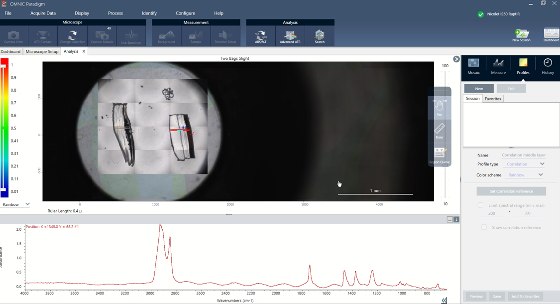
Image credit: Thermo Scientific - Chemical Analysis
Paint coatings are another form of micro layers. Automotives have different paint coatings; the top layer, for example, is a clear coat that offers shiny gloss and protects it from oxidation.
Some of these packages need to be durable when you ship them. Some handlers at different shipping sites throw it on the back of the truck, so you do not want your packages ripping.
It is also important to be able to recycle or reuse the packages.
One example I like to give when it comes to layers and packaging is the ‘meals ready to eat’ (MREs) used by the military. They must be tough because they have to survive war and plane drops and keep food fresh for at least three years.
The drawback is that some of their materials are not recyclable, so they usually end up in landfills. Current research is trying to alleviate this using microlayer analysis.
The central microlayer application I will focus on is food, medication, or shipping packaging. There are different layers in packaging. For example, you have an outer layer such as PET that you can coat and print on.
There may also be a tie layer that combines two dissimilar layers. For example, some things do not combine easily as their chemistry differs. A tie layer can mix these two things together, with an oxygen layer sitting there and keeping oxygen from going in.
A seal layer, such as polyethylene, is also needed. Typically, producers take these polyethylene bags, run them through, seal them with heat, and then seal them together.
We can study a lot of this with microscopy. With the RaptIR, you can see different layers and overlays of the video and 2D images. You can clearly distinguish between each layer visibly and through the 2D image.
How important is sample preparation for microscopy?
Sample prep is essential in microscopy. I was always told the quality of your spectrum is proportional to the time you spend prepping samples. With laminates, a microtome is a really good tool, although not everyone can afford these.
I have different razor blades of different widths. I take the sample and lay it between two glass slides, exposing a portion of it on the top and taking a thin razor blade and cutting it right down. I cut off a little sliver and put it in a diamond compression cell. You put the other diamond cell on top and press it down. You need a thin piece for the IR to go through whilst not absorbing too much.
What does the OMNIC Paradigm software do?
The OMNIC Paradigm software is the fun part. With the OMNIC Paradigm software, the first thing that opens up is a dashboard. There are three main sections. The first section is where you set up your experimental parameters, and the second is where you store your data.
The third section is the workflow, which is more like an automated section where you set up a process and data collection.
The new management section gives you options for more specialized measurements.
Different experimental settings have various parameters depending on what you plan to run. If you are doing the same experiment over and over or the same handful of experiments repeatedly, you can preset these parameters.
In my typical transmission experiment, the aperture is an important feature since it is an experiment with spatial resolution. You change the aperture step size, height, and width when carrying out your experiment.
There are multiple ways to set up experiments. You can have it all automated, or you can do it manually.
Once you have the mosaic, you can set a background point. You would then draw out two area maps on the first and second samples. Collection time depends on the sample, but it can be set and left.
Can you provide an example of utilizing the OMNIC Paradigm for spatial resolution?
Take two random food packages. Perhaps you want to see if there is any difference between a generic type and a brand name. Right off the bat, you can see different layers. Each sample will have some layers associated with them, and the spectrum changes for each layer.
I can create and set a correlation profile if I find an interesting area. You can select a new profile, name it “top layer”, set it to correlation, and then see the chemical profile to view which samples have the highest correlation. Samples with similar spectrums are suggested as a similar material.
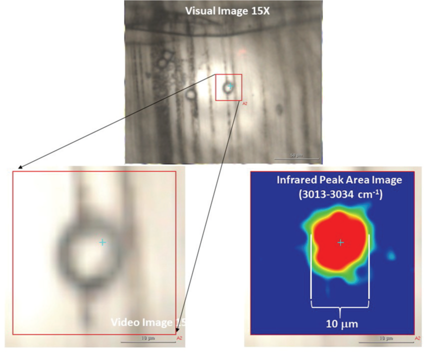
Image credit: Thermo Scientific - Chemical Analysis
One advantage of the OMNIC Paradigm Software is saving profiles for later use. For example, with common things like CH stretches and OH stretches. If you see an area with an OH stretch, the area can be set aside and saved as a baseline. You can then easily see that in two samples with an OH stretch in a similar area, the materials are relatively similar. Many samples have an OH stretch, so you might save it as a favorite for easy future use.
The ruler is another beneficial function of OMNIC Paradigm Software that shows the value of spatial resolution.
Please can you summarize the benefits of the Nicolet RaptIR FTIR Microscope?
The Nicolet RaptIR FTIR Microscope has new state-of-art features that can solve problems. It is automated for ease of use with an automated stage, objective, and focusing. There is improved security with database software and you can save multiple chemical profiles for future analysis of similar material.
You also have the ruler function to measure the width of micro layers and the ability to map multiple samples to make collecting data easier, more automated, and much faster. This is excellent for quality control, forensics purposes, and pharmaceuticals.
About Andrew Schmitz
Andrew Schmitz earned a B.S. in Physics and B.A. in Chemistry from Northwest Missouri State University. He received his Ph.D. in Chemical Physics from the University of Nevada-Reno researching spectroscopic probes via two-dimensional infrared. He also earned an MBA in conjunction with his Ph.D.
Andrew is currently an Application Scientist for the Nicolet FTIR spectroscopy product line at Thermo Fisher Scientific and works in the applications lab in Houston, TX.

This information has been sourced, reviewed and adapted from materials provided by Thermo Fisher Scientific – Chemical Analysis.
For more information on this source, please visit Thermo Fisher Scientific – Chemical Analysis.
Disclaimer: The views expressed here are those of the interviewee and do not necessarily represent the views of AZoM.com Limited (T/A) AZoNetwork, the owner and operator of this website. This disclaimer forms part of the Terms and Conditions of use of this website.