Analytical high-performance liquid chromatography (HPLC) is being extensively used as a method of identifying and quantitating molecular species present in a sample. HPLC systems include a range of online detectors including UV/Vis absorption, fluorescence, evaporative light scattering (ELS) and mass spectrometry (MS).
Differential refractive index detection (dRI) is also common, serving a large variety of HPLC applications ranging from small molecules to proteins, petroleum, polysaccharides and synthetic polymers.
Ultra-high performance liquid chromatography (UHPLC) is similar in applications and concept to HPLC but operates with smaller columns, packed tightly with smaller beads. This article presents several basic UHPLC analyses based upon an on-lined RI UHPLC detector. It can be observed from these examples that on-line UHPLC dRI detectors do not compromise UHPLC resolution.
The applications covered include column calibration, discrimination between eluting materials based on the ratio of UV:dRI, response linearity to different injected masses, detection of a non-UV- active impurity, and detection of a sample peak against the background of a solvent gradient.
Protein Fragment or Impurity
Bovine serum albumin (BSA) is a common reagent for calibration of protein quantitation and concentration measurements. Even though the monomeric content of BSA is as high as 97-98%, there is a small quantity of proteinaceous material in the solution that is not a BSA monomer. Most of the additional protein are irreversible aggregates of BSA (i.e., dimers and trimmers). These concentration-independent oligomers are analyzed and detected clearly by HPLC size-exclusion chromatography coupled to multi-angle light scattering (SEC-MALS).
High-resolution separation by means of UHPLC results in a new feature in BSA, a small peak eluting later than the primary monomer, shown in Figure 1.
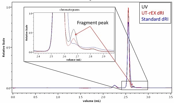
Figure 1. A minor impurity or fragment detected in BSA by UHPLC with a 300mm size-exclusion column and UV + UT-rEX RI. The different UV:RI ratio of the fragment relative to the monomer and dimer indicate a different primary composition.
The dRI chromatographic traces show a new peak. There are threefold sources of this peak, a misfolded BSA monomer, a fragment of BSA or a foreign molecule. Any variants of whole BSA molecules such as aggregates or miss-folded protein have the same composition ratio and hence have a constant UV:RI ratio; (e.g. the BSA dimer exhibits the same ratio as the monomer). The UV:RI ratio of the new peak is 60-70% higher than that of the monomer, implying that the new species is enriched in UV-active material relative to the monomer and, therefore, cannot be an aggregate or misfolded BSA. It must be foreign or a fragment.
Isomers
The interaction between a size-exclusion column and a macromolecule is mainly hydrodynamic in nature. Hence the elution time of any given species is governed by the shape and size of the molecule. Proteins having the same molar mass but different conformations interact a little differently with the column beads to elute at slightly different times. Standard HPLC size exclusion chromatographies, or UHPLC SEC using a short column (150mm), do not normally resolve the isomers unless they have vastly different shapes, (e.g., one is denatured).
Universal Detection and Column Calibration
Most detection techniques have solvent-dependent or specific sample limitations. UV or fluorescence analysis requires that the sample contain suitable UV-active chromophores and evaporative light scattering requires a volatile solvent. A partial list of samples compatible with the dRI is: proteins, carbohydrates, oligonucleotides, lipids, small-molecule drug compounds and oils. Figure 2 shows how the online UHPLC dRI detector was used in conjunction with a reverse-phase UHPLC column developed for the separating of sugars.
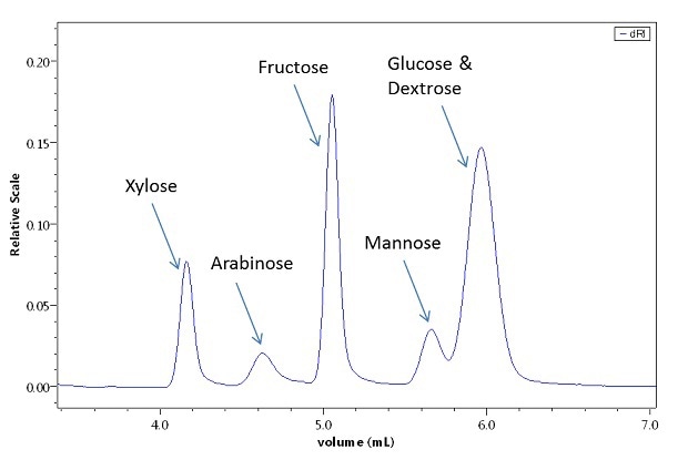
Figure 2. Sugar separation on a special UHPLC column, detected by the UT-rEX.
UV-Inactive Impurity Detection
Quality control of a UV-active substance, such as a protein, typically includes analytical UHPLC-SEC incorporating a UV detector. However, impurities present in the sample may not always contain chromophores and so will pass through undetected. In that case, adulterated samples will slip by QC tests without raising any warning flags.
Sample Detection against a Solvent-Gradient
Normally solvent gradients are accompanied by considerable changes in the refractive index of the mobile phase, overwhelming and saturating most on-line differential refractive index detectors. Whilst the preference is generally to use a different detection technology in gradient-based separations, sometimes RI may be the only option available. Figure 3 shows the analysis of a reverse phase acetonotrile/water gradient.
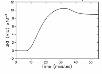
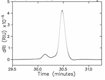
Figure 3. Analysis of a reverse phase acetonitrile/water gradient, 10% → 90%, by dRI. The sample is lysozyme. Top: raw data with 250 µg injection of lysozyme which appears as a small feature on a drastically changing RI signal. Bottom: after baseline subtraction, this time from a 25 µg injection of lysozyme (which is practically invisible against the gradient baseline). The early-eluting, small peak is unidentified.
Conclusion
Refractive index detection affords a number of important benefits to UPHLC. As a universal detector, an on-line UHPLC dRI detector (such as the Optilab® UT-rEX™ from Wyatt Technology) can be operated either stand-alone, as the sole on-line detector downstream of a UHPLC column, or in combination with another detector such as UV/Vis absorption.
It is possible to implement high data rate scan and low peak broadening to make sure that the improved resolution of UHPLC is not compromised. Stand alone detection mode is suitable for those samples that are not suitable to other measurement technologies, such as sugars in aqueous, non-volatile solutions, or proteins in solvents that absorb UV. The UV:RI ratio can also be analyzed to determine the composition of a conjugate molecule, (e.g. a protein carbohydrate biotherapeutic). This extra capability adds significant value to UHPLC analysis.
This content has been reprinted with permission from "The Benefits of Refractive Index Detection for UHPLC" by Dan Some, 2013. Asia Pacific Biotech News, Volume 17, No. 9, pg. 26-28.
About Wyatt Technology
With a long history of excellence in scientific instrumentation, Wyatt Technology is the recognized leader in innovative, quality light scattering instruments and software for determining the solution properties of macromolecules and nanoparticles in solution:
• Molar mass
• Size
• Charge or zeta potential
• Biomolecular interactions
• Conformation and composition
Wyatt provides solutions for in-line multi-angle static light scattering (SEC-MALS), field-flow fractionation (FFF-MALS), composition gradients (CG-MALS), high-throughput and traditional dynamic light scattering (DLS), electrophoretic mobility (ELS), differential refractometry and differential viscosity. Through a staff comprising 20% Ph.D. scientists and many more dedicated and experienced support personnel, Wyatt's aim is to delight the customer with the best training, customer support and service available in the industry.

This information has been sourced, reviewed and adapted from materials provided by Wyatt Technology.
For more information on this source, please visit Wyatt Technology.