In this interview, AZoM talks to Brent Nannenga, Ph.D., Assistant Professor at Arizona State University, about MicroED and its potential.
What is your background with electron microscopy? How long have you used EM in your work?
I first encountered electron microscopy when I started my postdoc with Tamir Gonen at the beginning of 2012. It was during my time there that we developed the MicroED (microcrystal electron diffraction) technique. Then, in 2015, I started my position at Arizona State; around the same time, they bought a Krios S/TEM. Since then we've been using MicroED and have gotten into single-particle analysis; we’re also hoping to eventually start doing cryo-tomography.
We use these techniques to answer biological questions, but we're also getting into materials science research and the study of small organic molecules. We try to tackle pretty much anything that anybody has a structural question about.
Can you give us the elevator pitch for your group’s current research?
There are really two sides to my lab. One side is protein engineering, really traditional protein engineering. Basically, we try to design proteins to do different things.
The other side focuses on cryo-EM (cryo-electron microscopy) methods, like MicroED, and their applications. The thing about Arizona State is that there's a lot of interesting cross-disciplinary research and we try to see how cryo-EM can potentially help with those projects. We try to modify existing methods or even develop new ones to help answer these interdisciplinary questions.
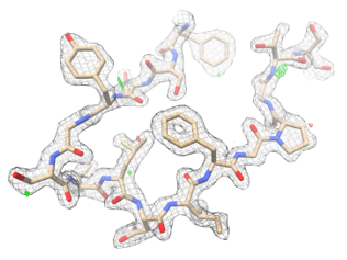
LCP-MicroED structure of proteinase K. Density map (wireframe) is superimposed with molecular structure (color).
For example, as I mentioned, we're getting more into materials science research, things like radiation sensors and inorganic materials. And, of course, there’s always new, interesting proteins and other biological systems.
What do you see in the future of cryo-EM? What needs to happen for us to get there?
That's a great question. I already think cryo-EM is fantastic, but continued development is critical. It's getting more user friendly, more robust with higher throughput; because of these improvements people from outside of electron microscopy are starting to see the benefit and really use cryo-EM.
In fact, I would say there seems to be a bit of a feeling like it's done. “Why do we need to keep developing it? We're already getting all these great structures!” But I think it's important to continue to push things forward. To find the next boundary and keep bringing this technique to more people.
To be perfectly honest, MicroED is working out really great. We're getting more collaborators than we know what to do with right now. So, we're often just doing "run of the mill" stuff. But from my perspective there's a lot of areas within this field that need more attention, and it's important we don't lose sight of that.
Is there any recent work from your lab that you would like to highlight?
Recently we’ve been focusing on using MicroED on samples that are in the viscous lipidic cubic phase (LCP). LCP has been successfully used to crystallize membrane proteins but it’s very hard to handle for MicroED. We’ve been able to look at a model soluble protein so far and are currently extending these sample preparation methods for membrane proteins in LCP. (Read more: Structure determination from lipidic cubic phase embedded microcrystals by MicroED) We’re also using MicroED in the area of materials science and for organic molecules to rapidly determine structures of beam sensitive materials.
About Brent Nannenga, Ph.D.
 Brent Nannenga, Ph.D. is an Assistant Professor at the Arizona State University School for Engineering of Matter, Transport and Energy as well as a Faculty Member at the Center for Applied Structural Discovery at ASU’s Biodesign Institute. We sat down with him at the 2019 American Crystallography Association Annual Meeting to discuss his background with EM and what he hopes to see in the future of the technique.
Disclaimer: The views expressed here are those of the interviewee and do not necessarily represent the views of AZoM.com Limited (T/A) AZoNetwork, the owner and operator of this website. This disclaimer forms part of the Terms and Conditions of use of this website.