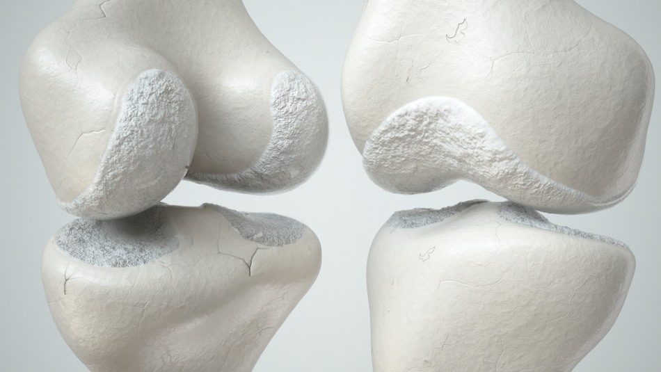
Image Credit: Crevis/Shutterstock.com
Some say 3D printing is a game-changer, but in the health and medical sector, it could also be a life-changer. The technology allows the creation of very precise, complex shapes, and has applications in printing cartilage, tissues and organs.
Three-dimensional bioprinting enables a more significant understanding of specific injuries and diseases, and could offer tailored treatments to meet individual patient needs. The technique involves depositing a bio-ink loaded with cells layer by layer, slowly building up tissue, organs or cartilage.
Creating Cartilage with Liquid Crystals
Cartilage is a complex porous structure; a compliant, elastic tissue that provides rubber-like padding to protect the extremities of long bones at the joints (areas such as the knee, ankle and elbow). It also forms part of the rib cage and intervertebral discs, ear, nose, and bronchial tubes, as well as many other body components.
Cartilage is soft enough to cushion joints, yet strong enough to resist compression. But it becomes damaged over time due to constant wear and tear, particularly in athletes.
Professor Chris Yakacki and colleagues from the University of Colorado Boulder, US, are the first to create cartilage using liquid crystal elastomers (LCE), known for their elasticity and ability to disperse high energy.
Although liquid crystals are found in everyday objects such as phone displays and polymers such as Kevlar, the challenge was to incorporate them into soft polymers where they would act as shock absorbers.
To create cartilage, the team employed a 3D printing technique called Digital Light Processing (DLP). They developed a honey-like liquid crystal resin which, when struck by ultraviolet light, formed new bonds in a succession of thin photopolymer layers. The resulting cured resin was a durable yet soft compliant elastomer and, when printed in a lattice structure, began to mimic cartilage.
The prototype spinal fusion cage showed 27 times greater strain-energy dissipation than those printed with commercially available photo-curable elastomer resins.
Yakacki is excited about the material’s possibility in the spine; previous attempts to mimic this structure have proved unsuccessful, but this method is precise and could match a person’s anatomy.
Bioprinting with Stem Cells
Bioprinting to manufacture human tissues and organs is the Holy Grail of research; not only could it signal an end to donor shortages, but it could also mean patients are treated with their own tissues.
Researchers from the Sahlgrenska Academy at the University of Gothenburg, Sweden, developed cartilage by printing stem cells onto a specially created scaffold. Cartilage cells from recent knee surgery patients were manipulated to become pluripotent, meaning they could develop into many different types of cells.
The stem cells, which were coated in nanocellulose to ensure they survived the process, were printed onto the scaffold. The cells then multiplied and were given growth factors to encourage them to differentiate into cartilage cells.
The 3D bio-printed structure closely resembled human cartilage, so much so that experienced surgeons could not tell the difference. When perfected, the cartilage could be 3D printed from the patient’s own stem cells to repair damaged cartilage or heal osteoarthritis.
But more work is necessary, as biocompatibility with the human body remains unclear. The structural material will need to degrade and be safely absorbed by the body, leaving only the cartilage.
Click here for more information on 3D printing technology
Bioprinting Bone to Heal Injuries
Researchers at Rice University and the University of Maryland, both in the US, created artificial tissues using 3D printing, which could help damaged bone or cartilage. Their work could help injuries – particularly those sustained in sports – in the knee, ankle, or elbow.
This collaboration aimed to mimic a tissue that gradually turns from cartilage (chondral tissue) at the surface, to bone. They developed a scaffold that reproduces the physical characteristics of an osteochondral lesion – a type of gap in the surface of cartilage in the talus (ankle) or tibia (shin) bone.
The team bio-printed a scaffold composed of custom mixtures of polymers for the cartilage part and ceramic for the bone part. The ceramic was embedded with pores to allow the patients’ cells and blood vessels to infiltrate the implant, meaning that the scaffold will eventually form part of the natural bone and cartilage, allowing osteochondral lesions to heal.
The next step is to determine how to print an osteochondral implant perfectly adapted to an individual patient, that will grow and fuse directly to bone and cartilage.
Realizing the Potential of 3D Printing in Biomedical Applications
Three-dimensional printing has revolutionized many industries, and adapting it to biomedical applications is likely to have the same effect in healthcare settings.
While more research is necessary, particularly into whether the materials used in the process are biocompatible, the future or limb repair is promising: from tailored scaffolds to mimic bone and cartilage, to the increasing prevalence of bio-printed tissues – it may even herald a new era of limb replacement technology.
References and Further Reading
New limbs could be built from scratch as scientists create cartilage with 3D printer. [Online] Metro. Available at: https://metro.co.uk/2020/06/08/new-limbs-built-scratch-scientists-create-cartilage-3d-printer-12820981 (Accessed 11th June 2020).
Motivans, E. (2019) Human Cartilage has been successfully 3D printed. [Online] ZME Science. Available at: https://www.zmescience.com/medicine/human-cartilage-successfully-3d-printed/ (Accessed 11th June 2020).
Carlota V. (2019) Bioprinted artificial tissue to heal bone and cartilage injuries. [Online] 3D natives. Available at: https://www.3dnatives.com/en/bioprinted-artificial-tissues-090420195/ (Accessed 11th June 2020).
Nguyen D et al. (2017) Cartilage Tissue Engineering by the 3D Bioprinting of iPS Cells in a Nanocellulose/Alginate Bioink. Nature Scientific Reports. https://www.nature.com/articles/s41598-017-00690-y (Accessed 12th June 2020).
Disclaimer: The views expressed here are those of the author expressed in their private capacity and do not necessarily represent the views of AZoM.com Limited T/A AZoNetwork the owner and operator of this website. This disclaimer forms part of the Terms and conditions of use of this website.