AZoMaterials speaks with Dr. Craig Johnson, Director of Research Core Facilities, and Dr. Kate Vanderburgh, Scanning Electron Microscope (SEM) and X-Ray Microscopy Manager, about the Materials Characterization Core (MCC) facility at Drexel University.
Tell us about Drexel University and the MCC facility.
Deep within the heart of a diverse city known for everything from the Liberty Bell to Cheesesteaks, resides a world-class R1 institution that is pushing the boundaries of educational success to new heights. At Drexel University in Philadelphia, they say it is ‘where academics meet the real-world’ and this is demonstrated through the use of not only putting students on the cutting-edge of innovation, but also ensuring their road to success post-graduation.
The founder of Drexel University, Anthony J. Drexel, once said, “I know the world is going to change, and therefore, the university must change with it.”
This statement resonates throughout the departments and curriculum, especially in the Materials Characterization Core (MCC) facility, which utilizes ever-evolving technologies to further scientific research and guide users on their path to becoming experts.
To achieve this mission of innovation and discovery, the MCC facility provides Drexel University faculty, staff, and students as well as external users with innovative materials characterization instrumentation, expertise, and training.
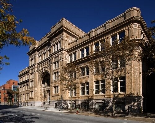
Image Credit: Drexel University
How does Dr. Craig Johnson and the MCC stay at the forefront of materials science research?
Dr. Craig Johnson has been involved with electron microscopy for over 23 years. Throughout this time, he has recognized many improvements and changes within imaging instrumentation.
To stay competitive and on the cutting-edge of materials science research, his team focuses on bridging imaging with analysis for applications in areas that encompass polymers, batteries, nanoparticles, and more.
However, as indicated by Drexel's founder that change is constant, the MCC's instrumentation must continually advance and evolve to maintain pace with the perpetual change of the scientific field.
How has the technology at the MCC evolved over the years?
The MCC was established around 2004 under a slightly different name and set of instruments than today.
Back then, it was primarily a collection of multiple systems, such as a FEI-Phillips (now Thermo Fisher Scientific) XL-30 Scanning Electron Microscope (SEM) and an older X-ray diffraction (XRD) system under one roof to form a technology instrument core that could better serve growing needs at the university.
Around 2008, the MCC continued to expand and over the next 5-6 years successfully acquired a FEI Strata DualBeam Focused Ion Beam Scanning Electron Microscopy (FIB-SEM) as well as multiple Transmission Electron Microscopes (TEM), XRD tools, NanoCT, and supporting equipment.
The MCC, due to its diversity of applications and multifaceted expertise, was able to successfully win multiple NSF-MRI awards that ensured the facility stayed up to date with current technology in its most heavily used areas.
In their most recent award, over 25 senior investigators were involved and they replaced the aging XL-30 with a modern Thermo Fisher Scientific Apreo 2 SEM.
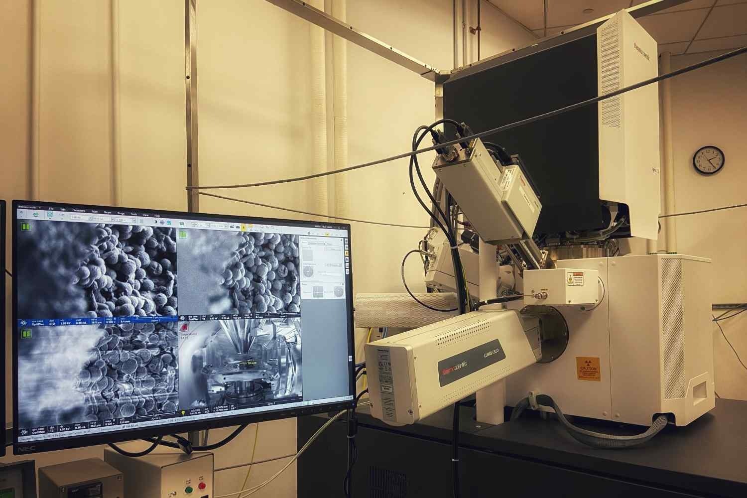
Image Credit: Drexel University
Why did Dr. Craig Johnson choose the Thermo Fisher Scientific Apreo 2 SEM?
The Thermo Fisher Scientific Apreo 2 SEM has many advantageous features. With high contrast and resolution, the Apero delivers high-performance imaging, even at long working distances.
It is compatible with various sample types and offers quick and easy sample loading. Thermo Scientific FLASH technology helps to automate the image fine-tuning processes and ChemiSEM technology provides live quantitative energy dispersive spectroscopy (EDS).
Dr. Craig Johnson knew that this system would open new doors for users to explore and correlate data from imaging, elemental, and in-situ studies across multiple platforms to shed light (as well as electrons) on some of today’s most challenging hypotheses.
Another aspect that ultimately led Dr. Johnson to choose the Apreo over similar systems was the user interface itself. Due to its intuitive and easy to understand layout, it enables efficient high-quality data aquisition by users from all experience levels.
One of the aspects that ultimately led us to choose the Apreo was the user interface itself… there isn’t a huge learning curve and the overall energy barrier to training is pretty low.
Dr. Vanderburgh, Scanning Electron Microscope (SEM) and X-Ray Microscopy Manager, Drexel University
How does the MCC enable scientific advancement?
Dr. Kate Vanderburgh leverages her material science expertise to educate users from diverse backgrounds on instrumentation so that they can solve complicated research questions and become independent, well-trained operators.
The main philosophy of the Dr. Vanderburgh is to train users on how to use the different instruments so that they become experts on not only their samples, but also the instruments themselves.
From the discovery of MXene materials at Drexel in 2011 to pushing the boundaries of today’s battery technologies, the MCC is a focal point enabling scientific advancement.
How has the Thermo Fisher Scientific Apreo 2 SEM contributed to the MCC’s mission of scientific advancement?
The new Apreo 2 SEM serves a very diverse group of researchers who are interested in all kinds of different materials.
The Apreo 2 has many bells and whistles for all facets of imaging and analysis. From beam deceleration and low-vac mode to Electron Backscatter Diffraction (EBSD), in-situ, and a CleanConnect sample transfer system, the instrument is primed for multiple research studies.
It is their 'go-to' SEM for demonstrations and challenging projects.
The microscope renewed what we were doing in the SEM space… for our core facility, this was the best thing that could happen.
Dr. Johnson, Director of Research Core Facilities, Drexel University
What educational programs and collaborations does Drexel’s MCC offer?
The MCC interacts with students, faculty, and staff at Drexel as well as extends its services to other institutions and industrial partners. At the foundation of their collaborative spirit is the understanding that properly training users is essential to education and advancement of science.
The MCC offers multiple programs for users, including 'full user training' and 'assisted use'. It regularly works with the University of Pennsylvania, Thomas Jefferson University, Villanova University, and others.
If readers have a project in mind, they should not hesitate to contact MCC regarding a collaboration.
The MCC is also heavily involved in outreach education, Women in STEM, and Drexel’s ‘Materials Science Days,’ which are typically held before Superbowl Sunday.
During these open exploration and education-packed days, the Apreo 2 SEM is used to teach students and the community about science and materials from the perspective of the nano-world.
The MCC feels that small things (even nano-sized) make significant differences, and teaching others about science is at the heart of Drexel’s mission.
What is next for the MCC?
The MCC’s team is often involved with the local Philadelphia Microscopy Society meetings, so look for society updates and future meetings. Also, the team at Drexel plans to hold workshops centered around electron microscopy in 2024.
The MCC will continue its training mission and enable the next generation of microscopists to help promote innovation and discovery. If you are interested in working with the talented team and advanced instrumentation, be sure to contact Dr. Johnson or Dr. Vanderburgh.
About Dr. Craig Johnson
Dr. Craig Johnson began his research career as a graduate student at Arizona State University working with transmission electron microscopes. After that he worked at Brookhaven National Lab in Long Island, NY then spent a year abroad as a post-doctoral researcher at a national lab in Toulouse, France. He joined Drexel University in 2008 to begin a transmission electron microscope program in the Centralized Research Facility, which has since been renamed the Materials Characterization Core (MCC). He quickly expanded to include a second transmission electron microscope and a focused ion beam scanning electron microscope. He took over as Director of Operations in 2017 and implemented many changes to keep the facility modern. He now holds the role of Director of Research Core Facilities. In his spare time, he loves fly fishing and dreams of fishing for giant trevally on a remote atoll in the Indian Ocean.
About Dr. Kate Vanderburgh
Hailing from New Jersey, Dr. Vanderburgh received her undergraduate degree in chemical engineering from Stevens Institute of Technology and a PhD in Materials Science from Vanderbilt University. Dr. Vanderburgh completed a post-doctoral appointment at Lawrence Livermore National Laboratory in California. She then joined the Drexel University team in 2021 as the MCC Research Instrumentation Specialist, managing the scanning electron microscopy and sample preparation tools. In her spare time, Kate loves hiking and counts the 215-mile John Muir Trail through the Sierra Nevada mountains of California as one of her accomplishments.

This information has been sourced, reviewed and adapted from materials provided by Thermo Fisher Scientific – Electron Microscopy Solutions.
For more information on this source, please visit Thermo Fisher Scientific – Electron Microscopy Solutions for Materials Science.
Disclaimer: The views expressed here are those of the interviewee and do not necessarily represent the views of AZoM.com Limited (T/A) AZoNetwork, the owner and operator of this website. This disclaimer forms part of the Terms and Conditions of use of this website.