In this interview, industry experts explain how Photothermal Stimulated Raman (PT-SRS) microscopy advances chemical imaging by combining Raman sensitivity with photothermal detection for faster, deeper, and label-free molecular analysis.
Could you explain what motivated the development of Photothermal Stimulated Raman, or PT-SRS, microscopy?
Prof. Cheng: Over the past 25 years, Coherent Raman Scattering microscopy, including CARS and SRS, has enabled high-speed chemical imaging. However, these techniques have limited sensitivity, typically around the millimolar level, which is insufficient for imaging biomolecules at physiologically relevant concentrations of nanomolar or micromolar levels. We wanted to overcome this limitation without relying on fluorescent or plasmonic labels. By revisiting the fundamental physics of the Stimulated Raman Scattering process, we realized that SRS produces measurable heat in the sample. Harnessing that heat through photothermal detection opened the door to higher sensitivity and more user-friendly, robust chemical imaging.
Within my research group we called this Stimulated Raman Photothermal (SRP), which has now been commercialized by Photothermal Spectroscopy Corp (PSC) and given the technique name of Photothermal Stimulated Raman Scattering (PT-SRS).
How does Photothermal SRS (PT-SRS) differ from conventional SRS or CARS microscopy?
Prof. Cheng: In conventional SRS, the signal depends on detecting very small intensity changes between the pump and Stokes beams, typically on the order of 10-5 (one in 100,000). In PT-SRS, we instead detect the small local temperature rise caused by the Raman process using a visible probe beam. This converts the thermal effect into an optical signal. The result is more than a 500-fold increase in modulation depth compared to SRS, providing much higher signal-to-noise ratios and greater sensitivity to low-concentration molecules.
What is the fundamental mechanism behind this photothermal effect?
Prof. Cheng: Stimulated Raman is a two-photon vibrational absorption process. When the pump and Stokes photons interact with a molecule, energy is deposited into vibrational modes, which then relax non-radiatively into heat. That heat changes the local refractive index and causes thermal expansion. We can detect these changes by focusing a separate visible probe beam slightly offset from the excitation volume, essentially measuring the thermal lensing effect. This makes the photothermal signal directly proportional to the amount of Raman absorption.
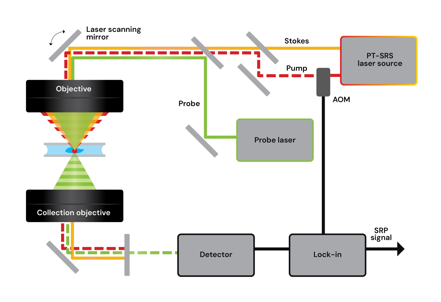
Image Credit: Photothermal Spectroscopy Corp.
How did your earlier research lead to the development of today’s PT-SRS microscopy?
Prof. Cheng: The idea traces back to an observation we made around 20 years ago while studying photodamage in CARS microscopy. We noticed that damage occurred more rapidly when the laser frequency matched a Raman resonance, which hinted that vibrational absorption was converting light into heat. At the time, that was an unwanted side effect, but it revealed an opportunity. If that energy deposition could be controlled and measured, it could form the basis of a new imaging contrast. Years later, building on our work in mid-infrared photothermal microscopy that later became the mIRage product (based on Optical Photothermal Infrared (O-PTIR) spectroscopy), we confirmed that the same concept could be applied to Raman excitation, giving rise to Stimulated Raman Photothermal (SRP) microscopy, now termed PT-SRS by PSC.
How does the sensitivity and resolution of PT-SRS compare to conventional SRS?
Prof. Cheng: The difference is significant. In our measurements, the modulation depth for pure DMSO was more than 500 times stronger with PT-SRS than with SRS. This means we can detect much smaller amounts of material while using similar laser powers. The spatial resolution is also improved because the photothermal signal originates from a very confined volume where all the beams overlap, reaching around 220nm laterally. That combination of sensitivity and spatial precision makes it possible to explore sub-cellular structures and complex tissue environments more clearly.
Beyond sensitivity, what makes PT-SRS microscopy easier or more practical to use?
Prof. Cheng: One of the appealing aspects of PT-SRS is its experimental flexibility. Because the signal is detected through a separate visible probe beam, the system can tolerate a wider range of optical configurations. Researchers can use longer working distance objectives and even simpler air-based optics without losing much signal quality. The detection scheme is also more forgiving of laser noise, which often limits conventional SRS. This makes PT-SRS a more robust approach for imaging live cells, thick tissues, or samples where environmental control is important. Another advantage is that the same principle can be adapted for both forward and backward (epi) detection, making it suitable for non-transparent or in-situ samples such as pharmaceutical formulations or biological tissues, something very difficult to perform with traditional SRS.
How is this technology implemented in the new stRAMos microscope?
Dr. Prater: The stRAMos Photothermal Stimulated Raman Microscope integrates this PT-SRS detection concept into a robust and easy-to-use instrument. It combines three co-linear beams, a pump, a Stokes, and a visible probe, and detects modulations in the probe intensity caused by local heating. Because we measure the probe beam, we are largely immune to noise from the excitation lasers. The system provides 10x higher sensitivity than conventional SRS, improved spatial resolution to below 300nm, and supports faster hyperspectral (through faster laser tuning and higher sensitivity) within a robust, align-free turnkey system, ideal for live-cell imaging, tissue imaging and other applications.
What type of laser technology enables such fast and sensitive imaging?
Dr. Prater: A major factor is the use of modern fiber-based SRS sources that can tune smoothly and rapidly across a wide spectral range. The laser we use can cover Raman shifts from about 3,100 to 700cm-1, covering the fingerprint and high-wavenumber regions. It can tune wavelengths in just a few milliseconds so that we can collect hyperspectral data much faster than with older solid state laser systems, typically used in traditional SRS, which can take seconds or longer to tune per wavelength. Because the source is fiber-based and computer-controlled, it stays well aligned and stable during long experiments. This kind of laser development has transformed what is feasible for vibrational imaging, transforming SRS chemical imaging from an advanced user technique requiring strong laser and optics expertise with dedicated large optical tables and specialized rooms, to a simple walk-up chemical imaging system.
We believe this will greatly expand the utility and usability of SRS chemical imaging, helping realize its full potential as an advanced chemical imaging modality.
Can you describe some of the imaging applications demonstrated so far?
Dr. Prater: We have demonstrated several types of applications. In biology, we can watch lipid droplets moving inside live yeast cells or examine how metabolites distribute within tissue sections. In materials science, the same technique can distinguish different polymer beads or detect microplastics based on their Raman spectra. The method also works in aqueous environments, which makes it useful for studying live cells without special preparation. Essentially, anywhere that chemical composition and spatial context matter, PT-SRS can provide valuable insight.
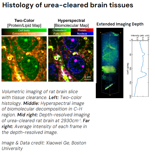
Image Credit: Ge, X., Zhu, Y., Sun, D. et al. Fiber laser based stimulated Raman photothermal microscopy towards a high-performance and user-friendly chemical imaging platform. PhotoniX 6, 35 (2025). https://doi.org/10.1186/s43074-025-00196-1
How large is the temperature increase during PT-SRS imaging, and does it risk sample damage?
Dr. Prater: The heating is extremely small and very localized. Each laser pulse raises the temperature in the focal volume by only a fraction of a degree to a few degrees Celsius, depending on the laser power and material properties. Importantly, this increase lasts only a few microseconds before dissipating, so the sample returns to its normal temperature almost immediately. That transient behavior is why we can image living systems safely, with little or no risk of thermal damage.
How does the visible probe beam contribute to the improved spatial resolution?
Dr. Prater: Resolution in optical microscopy is fundamentally linked to the wavelength of light. Conventional SRS uses two near-infrared beams, so its spatial resolution is diffraction-limited by those longer wavelengths. In PT-SRS, our 532nm probe beam nearly halves that wavelength, producing a smaller focal spot laterally and axially, generating a ~2x improvement in spatial resolution over traditional SRS. Furthermore, since the effective imaging resolution depends on the overlap of all three beams, the result is tighter confinement and finer spatial detail than even the diffraction limit of the probe alone.
Finally, can you give an overview of how the stRAMos platform integrates with other optical, photothermal or Raman modalities?
Dr. Mustafa Kansiz: The stRAMos Photothermal Stimulated Raman Microscope is part of a new versatile laser-scanning multimodal optical platform. Alongside PT-SRS, users can perform Optical Photothermal IR (O-PTIR), Spontaneous Raman, and fluorescence microscopy in wide-field and confocal modes. In this era of data-hungry Artificial Intelligence (AI), being able to provide many different correlative, multimodal data channels will no doubt contribute to the extraction of more useful and meaning data from the sample, leading to faster and more impactful scientific discoveries. We are also expanding the platform with the upcoming mIRage-HSi Microscope, which adds laser-scanning O-PTIR for >30x faster and more flexible correlative imaging. Together, these systems give researchers a unified solution for label-free, high-resolution chemical imaging across both infrared and Raman spectroscopies, whilst also allowing for traditional fluorescence imaging, thereby providing for the best of all modalities.
About Dr. Ji-Xin Cheng 
Professor Ji-Xin Cheng is the Moustakas Chair Professor of Photonics and Optoelectronics at Boston University and holds a PhD in Chemistry from the University of Science and Technology of China. He is the co-inventor of CARS microscopy and co-founder of Vibronix Inc. He has authored over 180 papers with 18,000+ citations and received multiple international awards for research excellence.
About Craig Prater 
Dr. Craig Prater, Chief Technical Officer at Photothermal Spectroscopy Corp., holds a PhD in Physics from the University of California, Santa Barbara. With over 140 publications and 9,200 citations, he received the 2023 Coblentz Society Williams-Wright Award for advancing vibrational spectroscopy and pioneering innovations in photothermal and nanoscale measurement technologies.
About Mustafa Kansiz 
Dr. Mustafa Kansiz, Director of Product Management and Marketing at Photothermal Spectroscopy Corp., holds a PhD from Monash University focused on FTIR spectroscopy and multivariate analysis. With over 25 years of experience in FTIR and Raman microscopy, he oversees product innovation, marketing strategy, and applications development across the company’s spectroscopy portfolio.

This information has been sourced, reviewed and adapted from materials provided by Photothermal Spectroscopy Corp.
For more information on this source, please visit Photothermal Spectroscopy Corp.
Disclaimer: The views expressed here are those of the interviewee and do not necessarily represent the views of AZoM.com Limited (T/A) AZoNetwork, the owner and operator of this website. This disclaimer forms part of the Terms and Conditions of use of this website.