In this interview, AZoMaterials speaks with Dr. Shan Zhou, Assistant Professor from South Dakota School of Mines, Dr. Grant A. Crawford, Professor and Director at the Advanced Materials Processing Lab, and Dr. Edward F. Duke, Director of the EMES Facility. They discuss how SEM and other advanced microscopy tools from Thermo Fisher Scientific support cutting-edge research, interdisciplinary collaboration, and STEM education.
Together, the faculty of South Dakota Mines illustrates how instruments like the Axia ChemiSEM, Helios DualBeam, Talos TEM, and Nexsa XPS form the foundation for next-generation workflows, from nanoscale imaging to materials processing and public outreach.
Can you please introduce yourselves and your roles at South Dakota Mines?
Dr. Shan Zhou: I’m currently an Assistant Professor in the Department of Nanoscience and Nanoengineering. My research centers around nanomaterials synthesis and characterization, especially using in-situ liquid-phase TEM to understand dynamic behaviors and self-assembly at the nanoscale.
I’m particularly interested in how nanostructures evolve in solution and how we can use those insights to design functional materials for applications in areas like drug delivery, biosensing, and energy storage. I also focus on integrating new technologies, such as automated TEM scripting, to streamline image acquisition and analysis.
Dr. Grant A. Crawford: I serve as a Professor in the Department of Materials and Metallurgical Engineering and direct the Advanced Materials Processing Laboratory. I lead a team of over 40 researchers, including graduate students, postdocs, and staff.
Our work emphasizes the structure–property relationships that underpin high-performance materials, especially in the context of advanced manufacturing such as additive manufacturing, advanced coatings, and joining processes. A critical element of our laboratory involves advanced characterization of materials, where we use tools such as the Helios DualBeam FIB-SEM, Axia ChemiSEM, Talos TEM, and Nexsa XPS systems. As an academic laboratory, we also emphasize undergraduate and graduate training, ensuring students gain hands-on expertise with state-of-the-art instruments.
Dr. Edward F. Duke: As Director of the EMES Facility, I oversee both the strategic development of our instrumentation suite and its integration into academic programs, outreach initiatives, and collaborative research.
Our facility supports the needs of multiple departments, and my focus is to ensure that the microscopy resources – including the Axia ChemiSEM, Talos TEM, and ancillary systems – are fully optimized for interdisciplinary research and education. I'm also heavily involved in community engagement, where we use instruments like the Axia SEM to spark interest in science among K-12 students and the public.
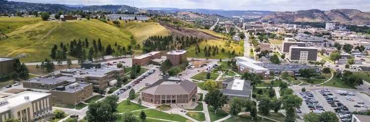
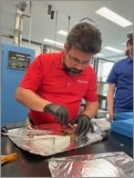
Image Credit: South Dakota Mines
How are you using Thermo Fisher Scientific equipment in your research programs?
Dr. Zhou: The centerpiece of my research is the Talos TEM equipped with a Hummingbird liquid-phase holder, which allows us to observe nanoparticles and their interactions in real time within a fluid environment. This setup enables us to capture critical self-assembly processes, nucleation events, and dynamic transformations that would otherwise be missed in dry-state imaging.
We’re also looking to expand into cryo-TEM to freeze and preserve intermediate states in nanoscale processes. By coupling this with Thermo Fisher’s automation capabilities, such as scripting for high-throughput imaging, we’re building a robust platform for statistically significant and reproducible nanomaterials characterization.
Dr. Crawford: Our lab makes extensive use of the Helios DualBeam FIB-SEM for cross-sectional imaging, site-specific milling, and sample preparation for TEM. The dual-beam configuration provides incredible precision in targeting features of interest, whether in advanced alloys, composites, or coatings. We also use the Axia ChemiSEM for broader morphological and topographical assessments, particularly for education and preliminary analysis.
The Nexsa XPS system supports our need for surface chemistry evaluation, especially in studying oxidation behavior and interface integrity in new material systems. These instruments form a cohesive workflow from fabrication to atomic-scale analysis.
Dr. Duke: The Axia ChemiSEM plays a multifaceted role in our facility. Beyond supporting research projects in geology, metallurgical engineering, and chemistry, it serves as an indispensable tool for teaching. Undergraduate students use it in lab courses to understand the surface morphology and failure mechanisms of materials.
We've also integrated it into outreach programs where prospective students, parents, and community members can view biological and engineered materials at high magnification, an experience that often leaves a lasting impression and fosters enthusiasm for STEM fields.
What makes the SEM platform, specifically the Axia SEM, so valuable for teaching and outreach?
Dr. Duke: The Axia ChemiSEM offers a rare combination of user-friendly design and advanced imaging capabilities, making it ideal for non-specialists and early-career researchers. Its intuitive interface shortens the learning curve, allowing students to quickly gain confidence in operating high-performance equipment. In educational settings, this is critical. We can guide students through hands-on sample preparation, imaging, and interpretation, all within a single lab period.
For outreach, the Axia ChemiSEM acts as a visual gateway into materials science. When visiting, students see the fine details of an insect wing or a metallic grain structure, which immediately captures their interest and opens the door to scientific inquiry.
How does SEM complement other techniques like TEM and AFM in your workflows?
Dr. Zhou: SEM serves as the first layer of screening in our multimodal workflows. It provides fast, reliable morphological imaging, which helps us identify features of interest before committing to time-intensive TEM experiments. In our work with nanostructures, we often use SEM to examine aggregation patterns or particle size distribution at the micro to nanoscale, and then we zoom into molecular interactions using high-resolution TEM.
We’re also integrating atomic force microscopy (AFM) for topographical and mechanical property mapping, creating a correlative pipeline that spans orders of magnitude in spatial resolution.
Dr. Crawford: For us, SEM is essential for visualizing surface features and assessing sample quality before precision FIB milling or further TEM analysis. SEM helps identify inclusions, porosity, and phase boundaries in metal alloys or ceramic composites, which is vital for guiding mechanical testing and modeling. We also use SEM in conjunction with energy-dispersive spectroscopy (EDS) to collect elemental maps that inform subsequent analyses.
The complementary nature of these techniques ensures that our research is both comprehensive and targeted.
How do these tools contribute to student training and workforce development?
Dr. Crawford: Hands-on access to advanced instrumentation is central to our philosophy. We train undergraduates, master's, and doctoral students to operate SEM, FIB-SEM, and XPS systems as part of their coursework and thesis research. Students don’t just generate data; they learn about instrument calibration, sample preparation, troubleshooting, and interpretation. These are critical skills that set them apart when applying for industry roles or national lab positions. Many of our alumni have gone on to work for aerospace, energy, and defense companies, and they often cite this practical experience as a differentiator.
Dr. Duke: We’ve built our microscopy facility to be inclusive and interdisciplinary. Whether it's a mechanical engineering student analyzing fatigue fractures or a chemistry student characterizing catalysts, they all leave with a deeper understanding of materials and the confidence to use high-end equipment. Moreover, students involved in outreach activities also become ambassadors for STEM, often demonstrating instruments like the Axia ChemiSEM to middle and high school students during campus visits.
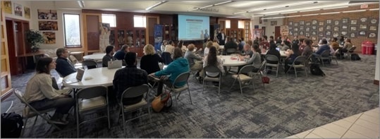
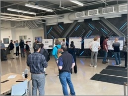
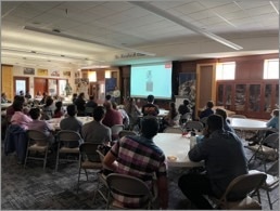
South Dakota School of Mines Workshop 2025. Image Credit: South Dakota School of Mines
Can you describe a recent research project that relied heavily on SEM or DualBeam instrumentation?
Dr. Crawford: One of our standout projects examined the microstructural integrity of additively manufactured metal alloys, particularly how build orientation and processing parameters influence grain structure and defect formation. SEM provided the first layer of analysis, revealing surface texture and porosity. We then used the Helios DualBeam system to create site-specific cross-sections, allowing us to visualize subsurface features like unmelted particles or interlayer voids.
The precision of FIB milling enabled us to prepare a TEM lamella from exactly the regions where we saw anomalies, giving us unparalleled insight into the causes of structural weaknesses.
How important is automation and scripting to your microscopy goals?
Dr. Zhou: Automation is critical for us because we’re collecting large datasets that require statistical rigor. With scripting, we can automate stage movement, image acquisition, and focus correction, which significantly reduces user bias and fatigue. This not only improves reproducibility but also allows us to explore a broader range of sample conditions.
In our in-situ experiments, automation ensures that we don’t miss transient events, such as sudden self-assembly transitions, which could be key to understanding the system’s behavior.
How have funding sources like the NSF MRI program supported your access to Thermo Fisher instrumentation?
Dr. Crawford: The NSF MRI program has been instrumental in helping us acquire the Talos TEM, Helios DualBeam, Axia ChemiSEM, and the Nexsa XPS system. These investments enable interdisciplinary collaboration and open new research avenues across departments.
By showing a strong record of student training, research productivity, and community outreach, we've been able to demonstrate the broader impact of these tools, which further supports future proposals and institutional development.
What role does SEM play in fostering collaboration with other institutions or industry?
Dr. Duke: Our open-access facility encourages partnerships across academia and industry. SEM often acts as the first step in collaborative projects–partners can quickly assess sample quality or damage mechanisms before proceeding to more specialized techniques. We've worked with mining companies, biomedical startups, and national labs, providing imaging and analysis that feed directly into product development or regulatory compliance.
This flexibility has positioned SEM not just as a research tool but also as a driver of innovation and commercialization.
Looking forward, what new capabilities or trends in SEM are you most excited about?
Dr. Zhou: Correlative techniques, especially those combining SEM with in-situ liquid and cryo workflows, are incredibly exciting. Being able to freeze and visualize nanoscale processes in real time opens new frontiers in dynamic materials science.
Dr. Crawford: I’m looking forward to integrating AI and machine learning into SEM analysis. Automating feature recognition and defect classification can significantly reduce analysis time and improve throughput in industrial settings.
Dr. Duke: From an education and outreach perspective, I see SEM becoming even more accessible, not just in terms of interface design but also remote operation and cloud-based data sharing. These innovations will allow broader participation in scientific research and education, particularly for under-resourced institutions.
This information has been sourced, reviewed, and adapted from materials provided by Thermo Fisher Scientific – Electron Microscopy Solutions North America.
For more information on this source, please visit Thermo Fisher Scientific – Electron Microscopy Solutions North America.
Disclaimer: The views expressed here are those of the interviewee and do not necessarily represent the views of AZoM.com Limited (T/A) AZoNetwork, the owner and operator of this website. This disclaimer forms part of the Terms and Conditions of use of this website.