Scanning electron microscopes (SEMs) facilitate significant scientific advancements by enabling researchers to investigate the properties of biological specimens and materials. However, traditional SEMs often present substantial size and cost constraints, limiting accessibility for many laboratories.
With technological advancements, there has been a growing demand for more compact yet powerful SEMs, particularly among laboratories seeking to enhance their imaging capabilities without the necessity of a large, dedicated space.
The Hitachi FlexSEM II effectively addresses this demand by combining tabletop models' convenience with full-scale systems' performance.
This versatile SEM, suitable for tabletop and floor-standing configurations, provides high-resolution imaging, adaptable sample handling, and user-friendly operation within a compact and flexible design. It represents an excellent choice for laboratories aiming to integrate advanced microscopy into smaller spaces or upgrade existing imaging setups without compromising quality.
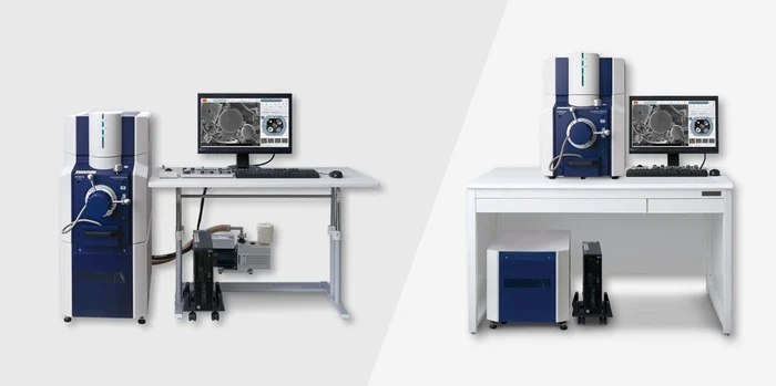
Figure 1. The FlexSEM II tabletop SEM. Image Credit: Hitachi High-Tech Europe
Why the FlexSEM II?
For many laboratories, the challenge lies in identifying an SEM that balances cost, size, and performance.
Convectional tabletop SEMs offer convenience but often lack the imaging capabilities required for detailed analysis. Conversely, full-scale SEMs perform well but entail significant space, cost, and operational demands.
The FlexSEM II solves these challenges by merging the advantages of both categories, delivering high-resolution imaging within a compact, lab-friendly design.
Designed for users of all experience levels, the intuitive interface and automation features streamline operation, improving accessibility for newcomers, while customizable settings address the needs of seasoned researchers. This flexibility enables laboratories to achieve superior imaging quality without the steep learning curve associated with more complex SEMs.
The FlexSEM II accommodates various applications, spanning materials science, biological research, and industrial quality control. The advanced imaging capabilities and adaptable sample handling boasted by the FlexSEM II make it ideal for labs aiming to broaden their analytical capabilities while adhering to budget and space constraints.

Figure 2. Super-absorbent polymer. No metal coating applied. Low-vacuum mode 30Pa, SE signal (UVD). Image Credit: Hitachi High-Tech Europe
Key Features
The FlexSEM II incorporates a range of advanced features within the compact SEM category. Below are the key characteristics that make it an optimal solution for laboratories seeking high-quality imaging within a smaller footprint.
Compact Design with Full-Scale Performance
Measuring just 45 cm in width and comprising a stackable electrical support unit alongside the SEM unit and an external vacuum pump, the FlexSEM II is sufficiently compact to fit on a workbench or function as a floor-standing unit.
This compactness does not compromise functionality; the system integrates a sophisticated vacuum system and high-performance electron optics.
Whether the user’s laboratory has spatial limitations or requires mobility, the FlexSEM II provides the flexibility to accommodate various installation setups and deliver the capabilities usually associated with larger SEMs.
High-Resolution Imaging Capabilities
With a secondary electron resolution of 4 nm at high vacuum, the FlexSEM II excels in producing detailed images for a diverse range of specimens.
The system offers an accelerating voltage range between 300 V and 20 kV, allowing researchers to optimize imaging conditions for different sample types and needs.
This versatility is particularly advantageous for laboratories that examine fine surface structures at low accelerating voltages or conduct detailed material analysis over various magnifications.

Figure 3. Toner particle, imaged at 5kV in high-vacuum mode with the Everhart-Thornley-SE detector. Magnification (polaroid base) is 20.000x. Image Credit: Hitachi High-Tech Europe
Adaptable Vacuum Modes
The FlexSEM II has dual vacuum modes: high vacuum for observing conductive specimens and low vacuum with adjustable chamber pressures up to 100 Pa for moist or non-conductive samples.
This adaptability minimizes the need for extensive sample preparation, facilitating efficient work with diverse materials, from advanced composites to biological tissues.
Advanced Detection System
In addition to an efficient high-vacuum secondary electron (SE) detector, the multi-mode backscatter electron (BSE) detection system enables high-sensitivity imaging across various vacuum conditions and accelerating voltages.
The FlexSEM is equipped with five BSE detectors, including options for material contrast and topographic imaging. Additionally, the optional low-vacuum secondary electron detector (UVD) improves imaging capabilities for non-conductive specimens.
A dual-channel display functionality allows for simultaneous viewing and recording of two image channels.
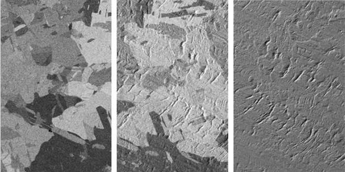
Figure 4. Examples of the different BSE imaging modes can be seen on the example of a rolled and briefly Ar ion beam polished Cu foil. Left: only grain orientation contrast without any topography is seen by the COMPO mode. Middle: by the 3D setup, sample topography (rolling traces) appear overlaid on the grain orientation contrast. Right :Pure topographical information is displayed by the TOPO mode. Image Credit: Hitachi High-Tech Europe
Flexibility and User-Friendly Operation
The FlexSEM II is designed with the user in mind, offering various features that facilitate ease of operation while delivering consistent, high-quality results. Flexible operation modes and an intuitive interface ensure that users of all experience levels can achieve excellent imaging performance.
Ease of Use
The graphical user interface is clearly structured and customizable, eliminating the need to recall functions hidden in sub-menus. A user-friendly operating knob panel provides a straightforward method to manage all major SEM functions.
Beginners can initiate their work with pre-set imaging conditions based on common applications, including surface topography and elemental analysis, allowing for the rapid capture of quality images without extensive technical knowledge.
For routine tasks, users can store custom settings in the recipe memory for rapid access to frequently utilized configurations.
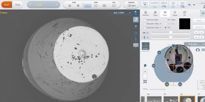
Figure 5. Basic FlexSEM graphical user interface, configured in single-screen mode. The SEM-Map function at the right side shows the colour navigation image, or optionally a second detector signal. At the lower right the thumbnail gallery of already recorded images is shown. Each image recording site can be revisited. Image Credit: Hitachi High-Tech Europe
Advanced users can control all imaging parameters completely, fine-tuning the system to meet their specific requirements.
Automated Functions to Streamline Workflows
The FlexSEM II includes standard and optional automated features that simplify and expedite common tasks.
The intelligent filament control optimizes filament saturation at each electron beam start, ensuring stable emission and prolonging the filament lifetime.
Standard functions such as auto-focus, auto-brightness, and contrast control help achieve optimal imaging conditions in seconds, significantly reducing the time required for manual adjustments.
Optional features like ZigZag-Capture, EM-Macro, and EM Flow Creator provide capabilities ranging from automatic multi-field image acquisition to customizable workflow programming for repetitive, complex imaging tasks.
Smart Navigation and Stage Control
Navigating samples is seamless with the FlexSEM's motorized specimen stage, which accommodates specimens up to 153 mm in diameter and 40 mm in height.
A trackball with a magnification-dependent response enables high-accuracy movement of the specimen stage in the X and Y directions.
The SEM MAP function integrates an optical camera view with the SEM interface, allowing users to quickly locate and focus on areas of interest. This navigation system ensures that even large or complex samples can be easily explored, enhancing productivity and ensuring accurate imaging results.
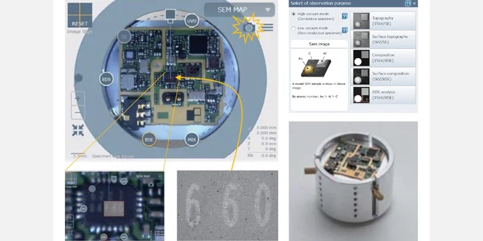
Figure 6. For easy orientation on specimen tables and convenient stage navigation, the FlexSEM incorporates a colour camera at the specimen chamber entrance. This camera acquires a high-resolution colour photo of any specimen loaded and makes this image available in the “SemMap” navigation window. Image Credit: Hitachi High-Tech Europe
Advanced Analytics and Optional Accessories
The FlexSEM II goes beyond standard imaging capabilities by providing analytical options and accessories that address a wide range of research needs.
EDS Integration for Elemental Analysis
For laboratories requiring detailed elemental analysis, the FlexSEM II supports integration with energy-dispersive X-ray spectroscopy (EDS) systems from leading providers such as Oxford Instruments and Bruker. These EDS detectors are compact and fully integrated within the FlexSEM housing.
These EDX systems enable precise detection of elemental composition, allowing for comprehensive material characterization. The option to select different detector sizes, up to 60 mm², ensures that the setup can be tailored to the analytical needs of each laboratory, whether for bulk sample analysis or high-resolution mapping of beam-sensitive materials.
3D Metrology and Image Processing
Enhance your analysis with the Hitachi Map 3D package, which incorporates powerful tools for roughness measurement, 3D surface reconstruction and particle, pore, or fiber analysis.
With data from the FlexSEM’s multi-detector system, the software generates detailed 3D surface models, enabling measurement of surface features and extraction of quantitative data such as step heights and texture parameters.
The package includes functions for stitching large, high-resolution images and color segmentation, facilitating the visualization of complex sample features.
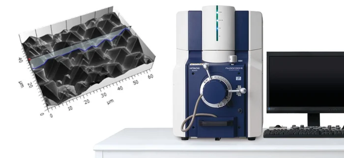
Figure 7. Together with a proprietary height calibration routine, the optional Hitachi Map3D Software will reconstruct an accurate 3D surface model of the inspected sample region. Image Credit: Hitachi High-Tech Europe
Cost Efficiency and Maintenance Advantages
The FlexSEM II provides advanced microscopy capabilities while addressing practical considerations regarding cost and maintenance. It represents a cost-effective choice for laboratories aiming to maximize their return on investment.
Low Maintenance Requirements
Engineered for user-friendly maintenance, the FlexSEM II allows most routine tasks to be performed in-house without the need for specialized technicians.
For instance, changing the tungsten filament takes just a few minutes due to video-animated user guidance and an easy-to-access electron gun design. Stable optics and automated alignment guarantee consistent performance, reducing the frequency of service calls and minimizing downtime.
Cost-Effective Solution
Operating costs are a critical consideration for every laboratory, and the FlexSEM II is designed with this in mind. Its self-maintainable components and efficient power requirements help minimize annual maintenance costs, often below €1,000.
This affordability, combined with high-resolution imaging capabilities, provides a compelling alternative to full-scale scanning electron microscopes, which incur significant ongoing maintenance expenses.
Energy Efficiency
The FlexSEM II operates on standard power and can be fully powered down when not used, restarting in less than five minutes. This energy-efficient approach allows laboratories to lower operational costs while supporting sustainability initiatives, making the FlexSEM an attractive choice for those seeking to reduce their environmental impact.
Conclusions
The Hitachi FlexSEM II delivers an ideal combination of compact design, high-resolution imaging, and advanced features that address the needs of modern laboratories.
It offers a practical solution for achieving high-quality microscopy without the space and cost requirements of traditional scanning electron microscopes.
With user-friendly operation, flexible analytical options, and low maintenance demands, the FlexSEM II is an accessible and versatile tool for a wide range of applications.

This information has been sourced, reviewed and adapted from materials provided by Hitachi High-Tech Europe.
For more information on this source, please visit Hitachi High-Tech Europe.