The key to Nanophoton's advanced technology employs is a line-shaped laser illumination combined with a two-dimensional CCD sensor to simultaneously capture 400 spectral data points in a single exposure. By utilizing galvanometer-based laser scanning, the RAMANtouch delivers high-speed, high-precision Raman imaging, efficiently mapping hundreds of thousands of pixels within minutes - all without relying on an EMCCD.
https://youtu.be/HerCE8T0n7k
This video shows the unique galvanoscanner of RAMANtouch in action.
RAMANtouch Key Features:
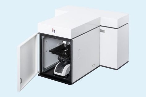
RAMANtouch microscope with laser class 1 safety housing. Image Credit: Bruker Optics
Laser Scanning via Galvanometers:
The RAMANtouch utilizes a galvanometric mirror for laser beam scanning, allowing the laser to move freely under the objective lens without shifting the sample stage. This method offers superior precision and speed compared to traditional table scanning. The perpendicular incidence of the laser beam across the observation plane enables specialized measurement modes:
- Laser Line Scanning
Scans the sample surface with high spectral and spatial resolution efficiently
- Point Scanning
Generates quick overview images by targeting areas with distinct Raman spectra.
- AreaFlash Measurement
"Flashes" the Raman laser line across the field of view to create an average spectrum of the entire area, facilitating ultra-fast measurements of large regions.
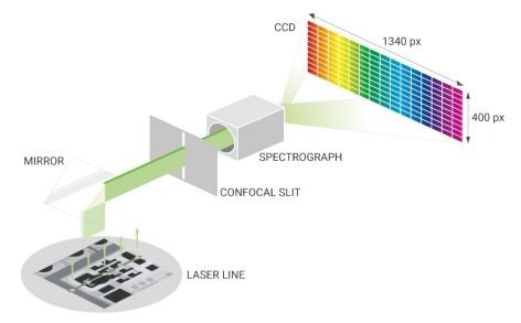
Image Credit: Bruker Optics
Laser Line Mode
- No emCCD required
- Ultra-fast Raman imaging by line illumination
- Homogeneous and deformation-free laser line over the entire field of view
https://youtu.be/CNDGDD3WPIE
Demonstration of Laser Line Mode for Raman Imaging. Video Credit: Bruker Optics
Laser Point Mode
- Up to 10 nm positioning accuracy
- Perfect focus spot, even at the edge of the field-of-view
- Hundreds of times faster than motorized stage
Demonstration of Laser Point Mode for Raman Imaging | Raman Microscopy | RAMANtouch RAMANwalk
Demonstration of Laser Point Mode for Raman Imaging. Video Credit: Bruker Optics
Exceptional 3D Raman Imaging Capabilities
The confocal optics of the RAMANtouch allow non-destructive analysis within samples. Using line illumination and a high-precision stage, it rapidly produces 3D Raman images of transparent samples, offering insights into internal structures and component distribution. For example, it can detect inside transparent fibers, such as bi-component PE (cladding) and PET (core) fibers, with acquisition times between 20 to 30 minutes.
Demonstration of 3D Raman Imaging | Raman Microscopy | RAMANtouch
3D Raman Image of two fibers. Video Credit: Bruker Optics
Raman Imaging Applications
Raman is used in battery research and industry to evaluate electrode stability and binder efficacy, which helps with performance and optimization.
Raman helps with performance improvement, fabrication optimization, and defect detection in silicon-semiconductor materials, guaranteeing effective device operation.
In life science, Raman helps with disease diagnosis, biomolecule research, cellular process comprehension, and biological sample structural analysis.
Raman microscopy is used in the pharmaceutical industry to analyze ingredient distribution, monitor formulations, and assess product quality.
Raman spectroscopy enables the analysis of crystal structure, phase identification, composition, and defects in inorganic materials and minerals.
Raman microscopy plays a key role in nano-carbon research and industry, enabling the analysis of graphene quality in CVD production and the characterization of carbon nanotubes for optimizing electronic applications.
In polymer and resin research and industry, Raman spectroscopy is used for material analysis and characterization, including evaluating the cure degree and resin distribution.
Raman spectroscopy is widely used in food science for quality control, contaminant detection, nutrient analysis, and studying food composition.
RAMANtouch Specifications
Source: Bruker Optics
| . |
. |
| Spatial Resolution |
350 nm in X, 500 nm in Y; 1 µm in Z |
| Objective Lenses |
5x, 10x, 20x, 50x, 100x |
| Spectral Resolution |
<0.9 cm-1 (depends on grating, up to 3 gratings available) |
| Stage Details |
30 * 30 * 35 mm XYZ-motorized stage |
| Calibration |
Auto-calibration based on standard lamp and sample |
| Alignment |
Auto-alignment of optical path |
| Laser Safety |
Laser safety class I door with interlock |