Image Credit: FLIR Systems
Generally, research and diagnostic biomedical applications require imagers with exceptional spatial resolution, precise color reproduction, enhanced sensitivity in low light conditions, and in a number of cases, a combination of all three factors to optimize data reliability.
Having the necessary cytology/cytogenetics camera, epifluorescence camera, histology camera, microscopy camera, etc., is key to offer a proper diagnosis in a clinical application or reliable data for the purpose of research. So how do you determine what machine vision camera is well-suited for your bioscience application?
In the sections that follow, several aspects to consider when choosing a machine vision camera for your biomedical and life science applications are outlined.
Application Specific Factors to Consider
Resolution and Color Accuracy
The appropriate resolution depends on the magnification of the structure of interest in the sample relative to the pixel size of the camera, i.e., high resolution in a microscopy application can be accomplished by a 2MP camera, a 25MP camera, or somewhere in between.
It is dependent on the structure of interest’s magnification in the sample by the optics corresponding to the size of the pixels on the camera. In order to choose the best camera options to reach the resolution desired, the determination of the size of the smallest structure in the sample to be resolved is necessary.
Then, multiply it by the lens magnifications in your optical system. This will generate the size of these structures when projected onto the camera sensor.
If the minimum size of the structure is 2.33 (Nyquist) times the size of a pixel on the camera sensor, then it should be possible to resolve that structure with the camera. For instance, if the size of these projected structures is ~8um, then a camera with 3.45um pixels can theoretically resolve such structures.
There are alternative methods when measuring resolution (e.g., line pairs), but this is a straightforward calculation to determine which suitable camera options to test.
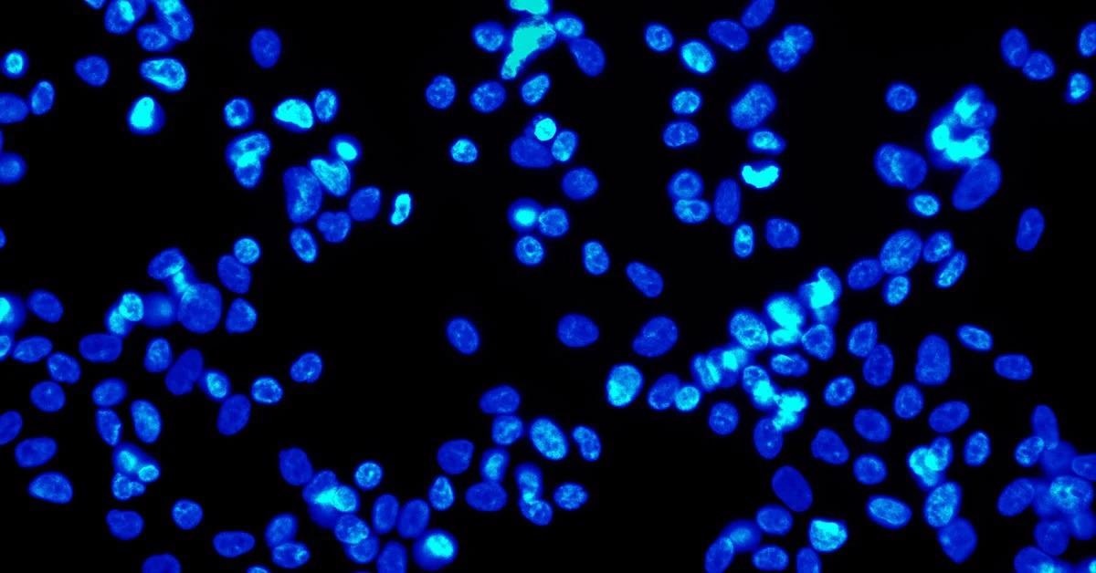
Imaging applications including histology, cytology, and cytogenetics operate across a wide range of white light (between ~400 nm and 700 nm) or utilize a selected wavelength within this range (for example, 565 nm).
If the specimens in these samples are living (or fixed), they can be exposed to bright light levels without the risk of killing the sample or stain fading. Under these conditions, the principal requirement for the camera is high resolution and color reproduction. To put it another way, low light sensitivity is not a key factor.
Sensitivity, Quantum Efficiency and Dynamic Range
Applications that call for the imaging of live specimens are met with the challenge of preventing overexposure of the sample. Too much light can kill the specimen or bleach fluorescent molecules. These applications generally use a method known as epifluorescence.
Epifluorescence techniques can be used on both fixed and live specimens. Acquisition of some specimens can be expensive, with others rare to find, and the process for creating samples can be costly in materials and labor.
Therefore, a system that protects the quality of the samples can help limit the recurring costs of these imaging applications.
To excite the sample, epifluorescence utilizes high energy wavelength that is filtered in order to emit a low energy wavelength. The low energy wavelength is filtered back to the camera.
Under these conditions, the main issue is sensitivity because this enables the use of less intense, damaging light on the sample. A camera with superior sensitivity can generate high quality images even when the light emission is low energy.
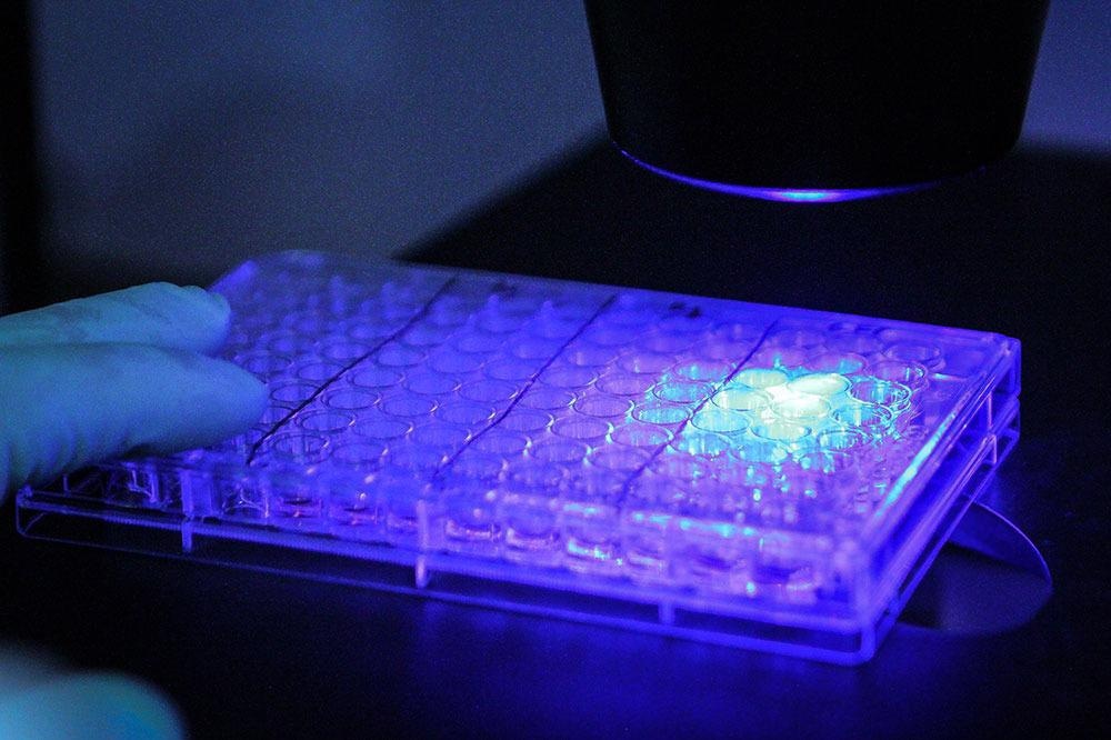
Image Credit: FLIR Systems
To determine the models with exceptional sensitivity that performs well under low light conditions, three specifications should be focused on: absolute sensitivity, quantum efficiency, and dynamic range.
Absolute sensitivity is the number of photons necessary to acquire a signal equivalent to the noise recorded by the sensor; the lower the number, the better.
Quantum efficiency is the percentage of photons transformed to electrons at a particular wavelength - here; a high number is better.
Dynamic range is the ratio of signal to noise, including temporal dark noise (the noise in the sensor when there is no signal). Here too, it is the higher, the better. For straightforward comparison, use the FLIR model selector to filter and determine the highest values.
Usually, monochrome models exhibit enhanced performance in low light in contrast to color equivalents.
To see the imaging performance details of a model, take a look at the comprehensive EMVA Imaging Performance document for the model on the FLIR website; these are accessible via the “Camera Resources” link for each camera family: Oryx Resources, Blackfly S USB Resources, Firefly Resources.
For further information on the EMVA imaging performance standard and how to cross-evaluate models for sensitivity, see How to Evaluate Camera Sensitivity.
Combination of Factors
For applications that utilize both white light and epifluorescence, consider camera models that are equipped with Sony’s new conversion gain feature, which has the capacity to optimize the sensor for increased sensitivity or high saturation capabilities.
High conversion gain is optimal for low light environments due to the minimization of read noise, which yields a low Absolute Sensitivity threshold ideal for identifying weak signals with short exposures.
Low conversion gain is the best for bright light conditions, as capacity of saturation is maximized, producing enhanced dynamic range. The maximum dynamic range will be inhibited by the 12-bit ADC.
For a list of models with conversation gain, please refer to the Machine Vision Sensor Review. For selection assistance with regards to the appropriate cameras for your particular application, get in touch with a FLIR machine vision expert.
Choosing the Right Cameras
When choosing a camera, a good starting point is selecting a newer CMOS sensor. Newer sensors tend to offer improved performance (and may be lower in price).
If the application in question necessitates the purchase of several cameras over several years (i.e., the continual manufacture of a diagnostic instrument), then it is crucial that a camera that is not at the end of its lifecycle is incorporated into the process - otherwise, additional costs will be incurred relative to prematurely designing-in a replacement camera.
FLIR produces over 200 variants of machine vision cameras that fit broadly into three camera families that utilize state-of-the-art CMOS sensors: Blackfly S, Oryx, and Firefly.
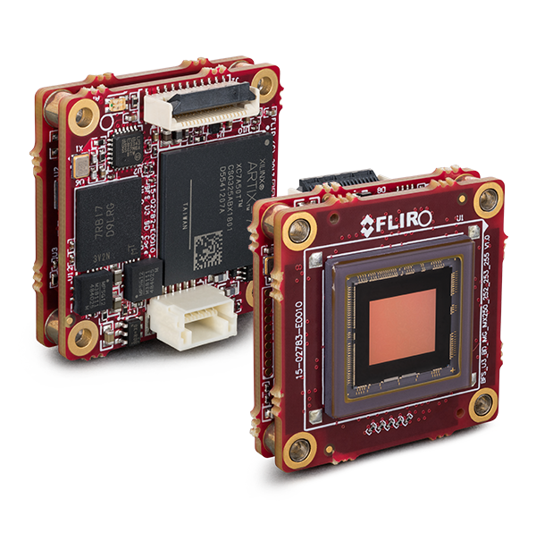
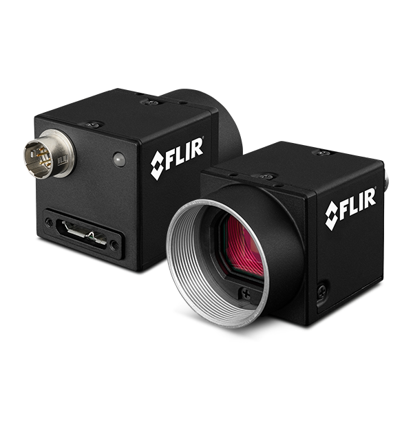
Image Credit: FLIR Systems
The Blackfly S camera family provides the widest range of sensors, form factors, and interfaces. With each model available in both USB3 and GigE variants, these cameras are exceptional in their versatility and easy to introduce to the design-in phase.
Board level Blackfly S versions are miniaturized versions of FLIR’s full featured cased range and are well-suited for embedded applications or those with space limitations. The diversity of features, excellent performance ratios and resolutions up to 24MP makes them a popular choice across biomedical and life science applications.
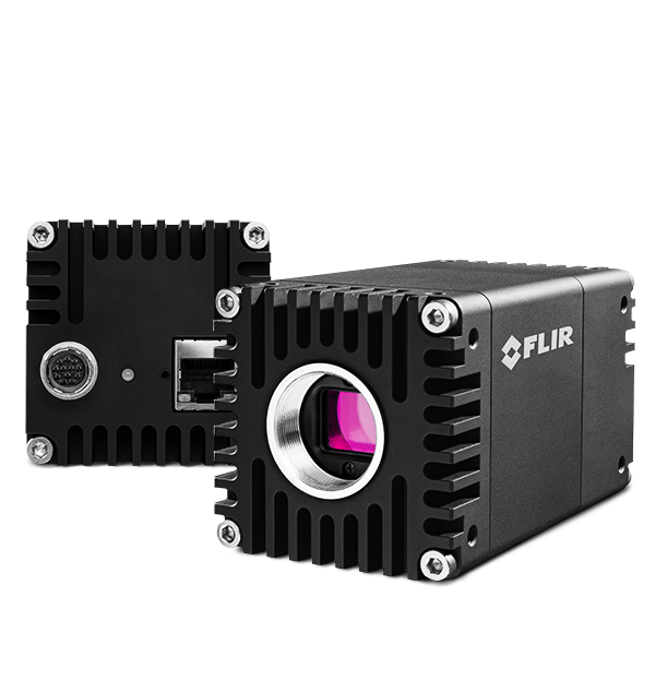
Image Credit: FLIR Systems
The Oryx camera family provides high resolution sensors paired with the rapid 10GigE interface, which allows for the capture of 4K resolution, 12-bit images at over 60FPS. Oryx’s 10GBASE-T interface is a widely deployed, proven standard that offers dependable image transfer at cable lengths in excess of 50 meters on inexpensive CAT6A or above 30 m on CAT5e.
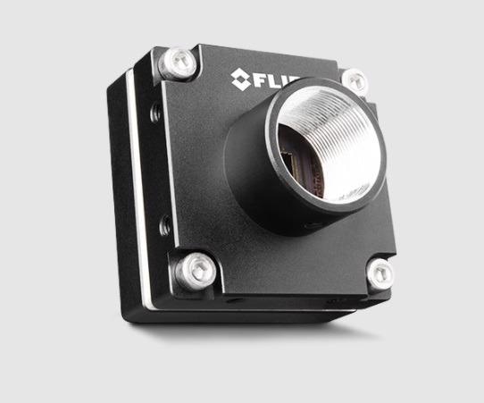
Image Credit: FLIR Systems
The Firefly camera family supplies a compact case form factor, lightweight and low power and price. The Firefly DL model also has the capacity to run a trained neural network that can be utilized for object detection or classification.
All FLIR machine vision color cameras come equipped with the capacity to customize color reproduction due to the availability of various white balancing options and the use of a novel color correction matrix, which is critical in biomedical imaging where color accuracy can mean different things depending on human visual analysis for diagnosis vs. a machine readable format for data accuracy.
For additional information on these features, see Using White Balance with Blackfly S and Spinnaker and Using Color Correction in Blackfly S and Oryx.
Moreover, the FLIR machine vision Blackfly S, Oryx, and Firefly camera families can be programmed and controlled using GenICam3 and the Spinnaker SDK, which has been developed from the ground-up with ease of future design modification and deployment in mind, this enables rapid application development and testing.
To narrow the selection of camera models, the FLIR website has a machine vision camera selector with multiple filter criteria available:
If you have any additional questions, FLIR’s machine vision experts are happy to help you select the right camera for your particular needs: click here to get in touch.

This information has been sourced, reviewed and adapted from materials provided by FLIR Systems.
For more information on this source, please visit FLIR Systems.