| Scientists at the Research Centre Julich in Germany have made individual oxygen atoms in perovskite materials directly visible using an electron microscope. Their success relies upon the development of a technique to correct the unavoidable aberrations in the microscope. In commercial instruments, these aberrations inevitably lead to blurred images - consequently no individual oxygen atoms can be recognised. The Julich scientists’ findings have been published in a recent issue of Science (7 February 2003). Pervskite Materials and Their Applications Ceramic materials based on oxides with a perovskite structure, that is a ratio of three oxygen atoms to every two metal atoms, such as barium and strontium titanate, play a major role in modern electronics. Their application is already widespread, for example, as chips in phonecards or paycards. Perovskites are also the base material for high-temperature superconductors, and will be needed in future microelectronics where they will be used in ultra thin films. One of the most important problems that needs to be overcome before perovskite materials can be used in this manner is to ensure the correct adjustment of the oxygen content of these oxides. This then has to be maintained through the large number of process steps in microelectronic device fabrication. ‘The oxygen content critically determines the electrical properties of the perovskite oxides,’ says Professor Knut Urban from the Julich institute of Solid State Research in Germany. ‘Since even the absence of a few oxygen atoms in the electrically active zones of the thin films would seriously impair their function, they must be fabricated with almost atomic precision.’ Transmission Electron Microscopy and Its Limitations In principle, transmission electron microscopy can be used to check if this atomic precision has actually been achieved. Consequently, researchers have been attempting to make the oxygen atoms in perovskite materials directly visible since the end of the 1980s so far without success. The basic principle of electron microscopy is quite simple - an electron beam penetrates a thin specimen. The outgoing electrons are guided by an electromagnetic lens system, which combines them into a greatly magnified image. However, for various reasons, particularly at high magnifications, you get distorted images and it is impossible to recognise individual oxygen atoms. Aberration Correction in Transmission Electron Microscopes The group headed by Knut Urban, however, has achieved a breakthrough. The scientists worked with an ‘aberration-corrected’ transmission electron microscope. The problem of ‘spherical aberration’ occurs in both optical and electron microscopes. Light or electron beams that pass through the lenses of the microscope close to the edge are deflected too strongly, resulting in an image that is blurred. With specially shaped magnetic lenses, however, the researchers are able to correct this previously unavoidable aberration. This step has proved to be extremely successful. It not only permits the individual oxygen atoms to be imaged for the first time, but also enables the oxygen content to be measured quantitatively in atomic dimensions. | 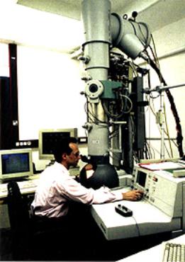
| | Figure 1. The silver section in the lower third of the electron microscope is the prototype with aberration corrected optics. | The Future for Aberration Correction in Electron Microscopes The scientists from Research Centre Julich developed the novel correction technique several years ago together with colleagues from the European Molecular Biology Laboratory (EMBL) in Heidelberg and Darmstadt University of Technology - thus creating the prototype of a completely new generation of microscopes. In the course of this year, the first commercial aberration-corrected electron microscopes will be available worldwide. Knut Urban is convinced of the importance of the aberration-corrected microscope. ‘This method will replace the classical type of high-resolution electron microscopy in many fields of materials science,’ he asserts. |