A light-responsive silicone adhesive lets flexible brain electrodes grip delicate tissue during recording and loosen when removal is needed.
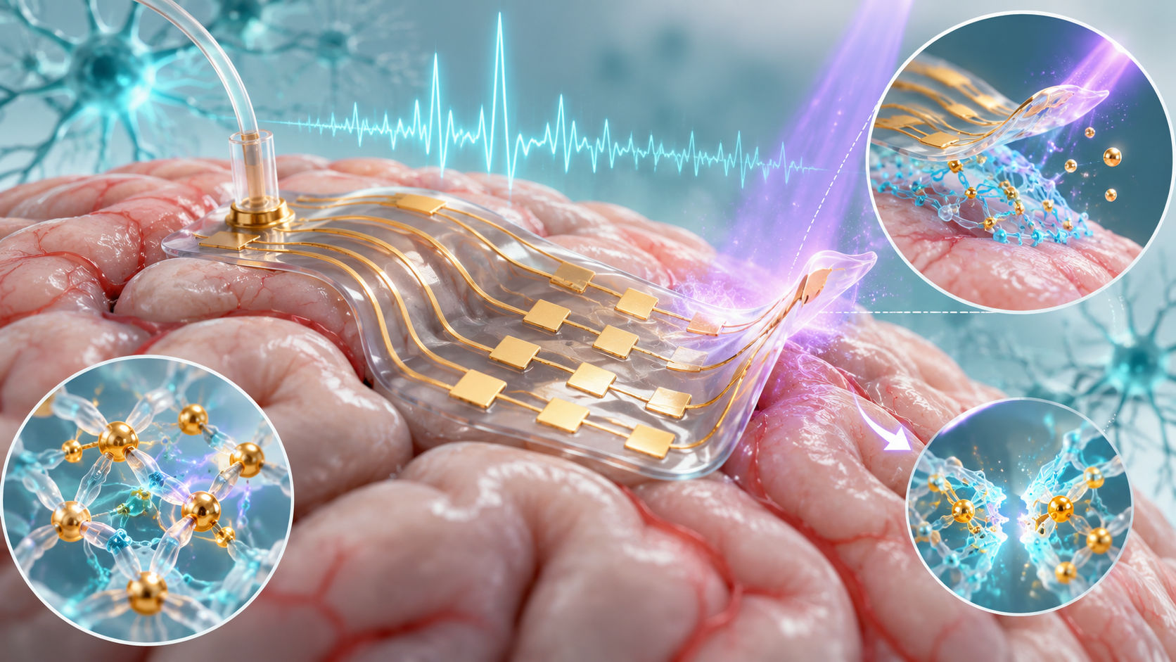
Paper: Light-regulated biomaterial interfaces enable flexible neural bioelectronics with stable adhesion and on-demand detachment for chronic electrophysiology. AI-generated abstract conceptual image created using ChatGPT/OpenAI
*Important notice: This news reports on an unedited version of an accepted paper and is awaiting final editing. Therefore, the paper should not be regarded as conclusive or treated as established information.
A recent study published in the journal npj Flexible Electronics introduced a light-responsive, self-healing elastomer called bPDMS@Fe for neural devices.
This material combines branched silicone networks with reversible iron-based coordination bonds, allowing it to adhere to metal surfaces with up to 310 N/m while conforming closely to complex neural tissue.
Under ultraviolet (UV) light, adhesion on glass fell from about 65 N/m to below 10 N/m after 15 minutes. In separate tissue tests, 60 seconds of 365 nm UV light at 16.09 ± 0.09 mW/cm² enabled clean detachment from porcine muscle, porcine skull, and mouse brain.
The challenges of neural interface adhesion
Long-term monitoring of chronic neurological conditions, such as epilepsy, can require neural interfaces capable of recording brain signals over extended periods. These devices must stay in close contact with brain tissue to accurately capture weak electrical signals. But existing materials struggle to balance firm attachment with safe removal.
Rigid electrodes can damage tissue and cause scar formation, while permanently adhered electrodes can concentrate peeling stress and raise the risk of cortical injury during removal.
Hydrogels can swell under physiological conditions and may leave gel deposits after removal.
Standard silicone materials, such as polydimethylsiloxane (PDMS), resist swelling in the body, but pristine PDMS has limited adhesion, which can allow electrodes to shift and introduce noise in recorded signals.
Synthesis and composition of bPDMS@Fe
To address these challenges, researchers developed bPDMS@Fe by adding reversible iron-based coordination bonds to water-resistant silicone. First, they used a UV-initiated thiol-ene click reaction to modify vinyl-terminated PDMS with 3-mercaptopropionic acid (MPA), introducing carboxyl groups into the silicone. Nuclear magnetic resonance (NMR) and Fourier-transform infrared (FTIR) spectroscopy confirmed the chemical changes.
Next, they dissolved bPDMS-COOH (carboxyl-functionalized branched PDMS) in tetrahydrofuran, then added an acetone solution containing ferric chloride (FeCl3). The resulting iron ions (Fe3+) formed reversible coordination bonds with the carboxyl groups, creating a reversibly crosslinked network with flexible and viscoelastic properties.
A nine-channel cortical electrode array was fabricated with several layers. A cured Sylgard 184 silicone base was coated with an approximately 50 μm bPDMS@Fe adhesive interface, followed by a 5 nm chromium (Cr) adhesion layer and a 100 nm gold (Au) conductive layer. A thin bPDMS@Fe layer supplied electrical insulation and tissue-facing adhesion. A guide cannula was co-implanted beside the array for targeted chemical delivery.
Detachment and self-healing mechanisms
Mechanical and spectroscopic tests showed that bPDMS@Fe combines tunable adhesion with autonomous self-healing capabilities.
In 90-degree peel tests, the material achieved adhesion strengths of 310 N/m on copper, 280 N/m on zinc, 210 N/m on iron, and 65 N/m on glass. It also adhered to biological tissues. On irregular isolated porcine brain tissue, the viscoelastic material relaxed its internal stress within 10 minutes, allowing it to conform to anatomical features.
Exposure to 365 nm UV light triggered on-demand detachment. The light reduced ferric ions (Fe3+) to ferrous ions (Fe2+) and partially decarboxylated the polymer, removing some carboxyl groups involved in coordination.
These changes weakened the coordination network and lowered the storage modulus, a measure related to the material's stiffness.
In the separate 60-second tissue test, the electrode detached without visible polymer residue or visible structural damage, and infrared measurements found no appreciable sustained temperature rise. The authors said the acute tissue tests did not rule out delayed effects.
The reversible coordination bonds also supported self-healing. After bisection, the elastomer recovered nearly 80% of its original mechanical performance within 90 minutes at room temperature.
When the elastomer severed gold conductive traces, it brought the separated pathways back into contact, restoring electrical continuity within seconds. Electrical recovery was also observed during stretch-release tests at strains of up to 20%. The gold layer itself did not heal.
Tissue compatibility and recording performance
In vivo tests on C57BL/6J mice assessed tissue response and recording performance. Brain histology at days 3, 7, and 28 showed transient, sparse astrocyte activation on day 3 and almost no detectable astrogliosis on days 7 and 28. Microglial density peaked at about 550 cells/mm² on day 3, fell to about 300 cells/mm² on day 7, and remained near that level at day 28. Three mice were examined at each histology time point.
In a separate group of three freely moving mice, the nine-channel array recorded focal seizures induced by 4-aminopyridine microinfusions, capturing synchronized spikes and sharp waves exceeding 700 μV.
Recordings were collected at days 7 and 14 after implantation, rather than day 28. The measured ictal-to-interictal signal contrast was lower at day 14 than at day 7, while multichannel recordings remained obtainable at both time points. The recording experiment did not include a parallel device-control group. The paper describes it as a feasibility experiment.
Applications beyond neural devices
Beyond neural implants, bPDMS@Fe may have uses in wearable sensors and other bioelectronic devices. In human skin tests, bPDMS@Fe electrodes showed smaller changes in skin-electrode impedance during prolonged contact. Both bPDMS@Fe and commercial gel electrodes produced identifiable ECG signals in dry and sweaty conditions.
During compression, stretching, and rotation, the commercial gel electrode partially delaminated, while the bPDMS@Fe electrode maintained close contact with the skin. Its switchable adhesion may also help position and remove bioelectronic interfaces when mechanical peeling could damage tissue.
What the study shows for neural interfaces
bPDMS@Fe combines light-controlled adhesion, mechanical flexibility, and self-healing in a flexible neural electrode system. The study included histological follow-up through day 28 in separate cohorts and neural recordings at days 7 and 14 in a three-mouse feasibility cohort. The results remain preclinical and feasibility-scale, but they support further study of removable flexible neural interfaces for electrophysiological monitoring.
Disclaimer: The views expressed here are those of the author expressed in their private capacity and do not necessarily represent the views of AZoM.com Limited T/A AZoNetwork the owner and operator of this website. This disclaimer forms part of the Terms and conditions of use of this website.
Source:
- Li, Z. et al. (2026). Light-regulated biomaterial interfaces enable flexible neural bioelectronics with stable adhesion and on-demand detachment for chronic electrophysiology. npj Flexible Electronics. DOI: 10.1038/s41528-026-00659-w, https://www.nature.com/articles/s41528-026-00659-w