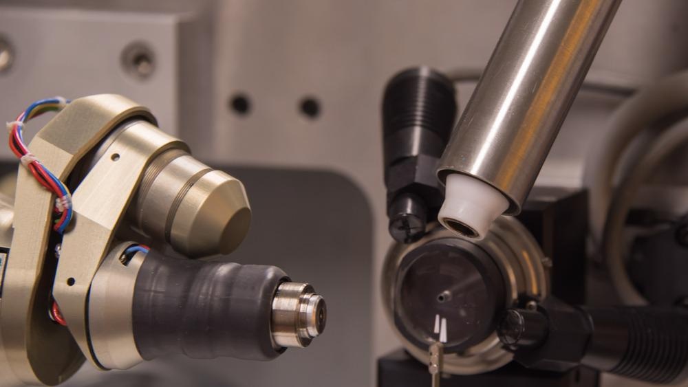
Image Credit: Gianmix/Shutterstock.com
Arizona State University researcher Petra Fromme has been awarded the 2021 Christian B. Anfinsen Award in honor of her contribution to the field of protein research. She has developed the use of ultra-high-speed x-ray crystallography to investigate protein structures in fine-grain detail and the shortest time durations. Here, we discuss how Fromme’s work is helping to move the field of protein research forward.
Fromme’s effort has advanced the scientific field in two key areas: streamlining pharmaceutical development for a range of human diseases, and alternative energy.
In her latest research, Fromme and her collaborators, including experts from various disciplines such as biology, chemistry, data science, engineering, materials science, and physics, established a cutting-edge method of serial femtosecond crystallography (SFX). Using its innovative method, the team visualized molecules in action at an unprecedented level of detail. As a result of this research, significant contributions have been made to the field of structural biology of proteins, particularly those involved in photosynthesis.
The Importance of Furthering Protein Research
Proteins are vital to the functioning of cells. Therefore, understanding more about their structure can help reveal essential information regarding their function and dynamics at a molecular level.
In comparison to DNA, which gives information in the form of letters that code for 20 different amino acids and proteins. A single protein can fold into an almost infinite number of three-dimensional configurations, which impacts how they interact with other molecules and ultimately impacts their function.
Gathering rich data on protein structure has the potential to deepen our knowledge of the roles proteins perform in our bodies. This knowledge is invaluable to the pharmaceutical industry as it can help compounds as candidate drug targets, speed up the development process, and create new therapeutic drugs for a wide range of diseases.
Visualizing Proteins
X-ray crystallography is one of the most relied upon and powerful techniques for visualizing protein structures. It involves the crystallization of a sample, which is then introduced to a beam of incident x-rays.
When the waves of the x-ray make contact with the sample, the light is diffracted, and the patterns of diffracted light are caught by a detector screen. Incredibly detailed three-dimensional images of the proteins are then produced by examining the sample in all possible orientations. With this method, researchers have looked at biological molecules in a way that has enhanced their understanding of their functioning and dynamics.
However, the method had not been without its disadvantages. As molecules exposed to the x-rays become destroyed because of the interaction, the method of crystalizing served to slow down this process. Unfortunately, this meant that only static pictures could be obtained. Fromme and her team developed a new version of x-ray crystallography to overcome this limitation. Known as serial femtosecond crystallography (SFX), Fromme’s radical new method exposes nanocrystals to incredibly short pulses of x-ray light (measured in femtoseconds), produced by x-ray free-electron lasers (XFELs).
Because the x-ray light pulses occur in femtoseconds, they can produce a diffraction pattern before the crystallized sample is destroyed in a technique known as “diffraction before destruction”. With this method, scientists can investigate a protein structure at room temperature and produce molecular movies, something which has revolutionized the field.
With their new innovative technique, Fromme and her collaborators were able to produce the world’s first molecular snapshots demonstrating the functioning of BlaC (an enzyme vital to antibiotic resistance) and Cytochrome c oxidase (fundamental to respiration). The team has also uncovered the detailed structures of numerous important proteins such as Flpp3 (responsible for the devastating effects of the tularemia-causing bacteria), angiotensin II (vital for regulating blood pressure), and DIPP-NH2 (which has a fundamental role in pain pathways).
It is likely that the SFX technique will continue to reveal more vital information on the structures of important proteins. Fromme believes that her team’s work will have a significant impact on the development of novel drugs as it will allow scientists to view drug targets in their active state, which will give more detail on their function.
Fromme also believes that the work will have a significant impact on the advancement of clean energy systems. She highlights that the technology can help gain a deeper understanding of how plants generate energy from a light source, information that is vital to developing efficient man-made systems that generate clean energy from renewable sources (solar power). Ultimately, there is an almost infinite number of ways in which SFX can benefit the scientific field given the huge importance of proteins on a wide range of functions.
References and Further Reading
Fromme's pioneering efforts in X-ray crystallography honored with prestigious Anfinsen Award. ASU News. Available at: https://news.asu.edu/20210412-frommes-pioneering-efforts-x-ray-crystallography-honored-prestigious-anfinsen-award
Gisriel, C., Fromme, P. and Martin-Garcia, J., 2020. Methods for Crystallization and Structural Determination of M-T7 Protein from Myxoma Virus. Methods in Molecular Biology, pp.125-162. https://asu.pure.elsevier.com/en/publications/methods-for-crystallization-and-structural-determination-of-m-t7-
Pandey, S., et al. 2019. Time-resolved serial femtosecond crystallography at the European XFEL. Nature Methods, 17(1), pp.73-78. https://pubmed.ncbi.nlm.nih.gov/31740816/
Disclaimer: The views expressed here are those of the author expressed in their private capacity and do not necessarily represent the views of AZoM.com Limited T/A AZoNetwork the owner and operator of this website. This disclaimer forms part of the Terms and conditions of use of this website.