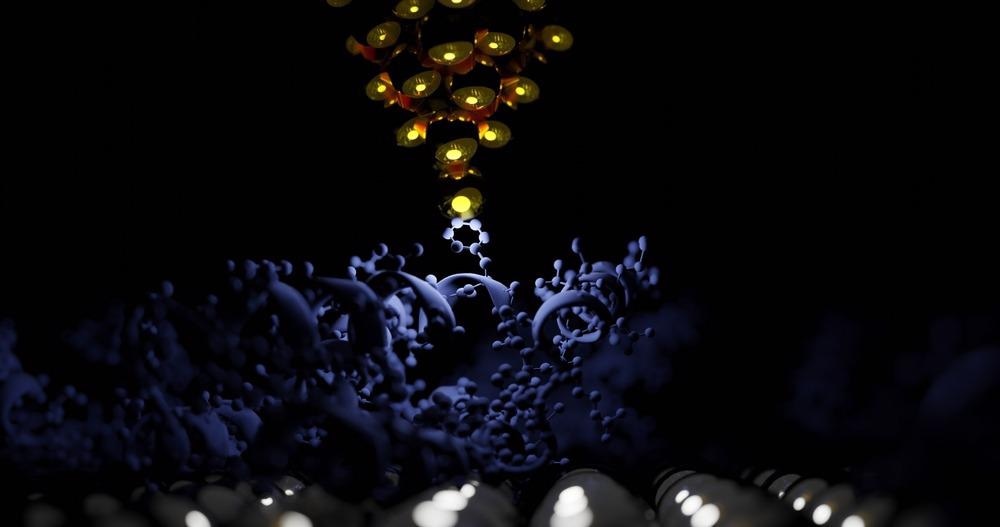
This article informs readers regarding the basics of atomic force microscopy, its uses for biological applications, and the different substrates used for AFM imaging of small biological objects.

Image Credit: sanjaya viraj bandara/Shutterstock.com
What is Atomic Force Microscopy?
Atomic force microscopy has been an effective and essential method utilized extensively for nanotechnology, physics, and biological applications. It is a surface examination technique and is widely used as an imaging method for obtaining high-resolution quality pictures of specific areas.
It is being used by nanotechnology researchers for the characterization of nanoparticles. The technology shapes a region by manipulating the interaction of stresses between a microscopic sensor and the area. The capacity to analyze interfaces with a high signal-to-noise level at sub-nanometer precision sparked the emergence of a number of AFM-related methods that utilize a range of instruments to selectively detect interactions and control materials.
Utilization and Advantages of AFM
AFM is a powerful method for analyzing nanomaterials and nanostructures. It gives qualitative as well as analytical data on a wide range of physical qualities such as dimensions, height, physical properties, and cleanliness. Many scientists from all around the world use AFM for materials research purposes.
AFM may be used to find nanoparticles, biochemical, biomechanics (modulus of elasticity, mobility, viscosity, resistance), and electromagnetic properties. AFM is less expensive, simpler to use, and can photograph at the atomic level when compared to other imaging methods.
Limitations of AMF
Even though AFM has been used by numerous businesses all over the world for a variety of purposes, certain restrictions remain a barrier to its widespread deployment. The sampling frequency of AFM is also a limitation. In the past, AFM could not scan images as fast as SEM, with an average scan requiring several minutes.
Because of the relatively modest scan rates during AFM scanning, metabolic destabilization in the image occurs often, making the AFM microscopy less ideal for establishing detailed measurements between topographic maps on the image processing. Image artifacts are conceivable, like with any other imaging method. They might be created by an unsuitable tip, a poor working atmosphere, or even the substance itself.
AFM Focused on Biological Applications
The establishment of an optical detecting device, followed by the construction of a hydraulic container, permitting scanning in buffer solution and so retaining the biological system's natural form, was the fundamental innovation that led to biological AFM. Multicellular organisms, cell membrane regions and protein complexes, DNA and RNA, and lipid coatings were among the biological items photographed shortly after the first commercially accessible atomic force microscope was introduced.
Although contact mode AFM is commonly used to analyze solid substrates, its use in soft biological systems necessitates professional knowledge of how to modulate the load exerted to the tip. Contact and dynamic mode AFM show topographical images beyond the resolution limit of traditional microscopic examination when applied to cellular systems.
What are Different Substrates Utilized for AMF?
AFM is useful in the macromolecular functionalization of small biological objects and membrane proteins, utilizing a substrate in the process. The most commonly utilized substrates involved are mica, silica, gold, silver, pyrolytic graphite, glass, Silicon dioxide, Zinc Oxide, Zinc Sulfide, and Calcium oxide. Apart from these, modified substrates have been developed by researchers for specific applications of AFM such as specially prepared substrates for DNA imaging, membrane analysis, etc.
Why is Mica the Most Utilized Substrate?
Mica, a negatively charged organic crystalline substance with varied chemical constitution and excellent hydrophilicity, is the most commonly utilized substrate. The most significant benefit of mica is that it may be readily split into molecularly flat surfaces with no crystalline discontinuities for hundreds of micrometers.
Mica is appropriate for single-molecule visualization of peptides, chromosomes, and carbohydrates, however it is susceptible to imaging artifacts because of a high surface charge and water vapor uptake from the air.
Graphite as Bio-substrate
Highly oriented pyrolytic graphite (HOPG), an unreactive artificial carbon crystalline substance, is another common substrate for biological AMF. It can be cleaved similarly to mica, however, it is less evenly flat due to many more crystalline layers per surface area. Furthermore, it is known that bare graphite causes structural changes in immobilized protein complexes.
More from AZoM: Analyzing Scale of Multi-Asperity Wear with AFM Techniques
Graphite has been changed via low-energy plasma or adhesion of long-chain aromatics, solvents, and fatty acids to increase its surface characteristics, making it appropriate for single-molecule photography of proteins and DNA. These changes, however, enhance the hardness of graphite.
Latest Advances
Researchers from Texas have compared the properties of various substrates such as mica, Si3N4, ZnO, CaF2, ZnS, and sapphire. It was found that mica, glass, Si3N4, Si, ZnO, and CaF2 have a flat texture that is ideal for AFM scanning of oligomers. Surfaces of CaF2 and ZnO, on the other hand, had lengthy, strictly delineated channels that reduced the integrity of the substrate but did not result in a significant increase in surface imperfection. Furthermore, CaF2, SnZ, Si, and sapphire, as well as KBr, exhibit extremely low to no IR background radiation within 800-1800 cm-1.
Because of the great solubility of the substrate in water, KBr has very little, if any, applicability for biological research. Furthermore, comparisons of Au-coated and bare Si, ZnS, and CaF2 substrates demonstrated that metal coating significantly increases the intensity of the spectral background. As a result, Au-coated substrates are unlikely to be useful for visualizing lipid-containing complexes with proteins.
However, as contrasted to the amplitude of the IR spectra acquired from amyloid clumps placed on bare Si, Au coating offers an enhancement to the IR signal by a factor of 7.
Future Prospect
During the forecast timeframe, between 2022 and 2029, the worldwide Atomic Force Microscopes (AFM) market is expected to grow at a rapid pace by the latest press release. For this purpose, customized substrates should be the focus that amplifies the imaging resolution for modern materials and nanotechnology. A lot of focus is being given to the production of substrates for fungi, viruses, and enzymatic research to help mankind progress and open new doorways.
References and Further Reading
Rizevsky, Stanislav, et al. 2022. Characterization of Substrates and Surface-Enhancement in Atomic Force Microscopy Infrared Analysis of Amyloid Aggregates. The Journal of Physical Chemistry C. Available at: https://pubs.acs.org/doi/10.1021/acs.jpcc.1c09643
Bobylev, Alexander G., et al. 2022. Amyloid aggregates of smooth-muscle titin impair cell adhesion. International journal of molecular sciences. 22(9). 4579. Available at: https://www.mdpi.com/1422-0067/22/9/4579
Hu, Bo, et al. 2022. Application of arbuscular mycorrhizal fungi for pharmaceuticals and personal care productions removal in constructed wetlands with different substrate. Journal of Cleaner Production. 130760. Available at: https://www.sciencedirect.com/science/article/pii/S0959652622003997?via%3Dihub
Akhatova, Farida, et al. 2022. Nanomechanical Atomic Force Microscopy to Probe Cellular Microplastics Uptake and Distribution. International journal of molecular sciences 23(2). 806. Available at: https://www.mdpi.com/1422-0067/23/2/806
Disclaimer: The views expressed here are those of the author expressed in their private capacity and do not necessarily represent the views of AZoM.com Limited T/A AZoNetwork the owner and operator of this website. This disclaimer forms part of the Terms and conditions of use of this website.