Researchers combine CMC-assisted electrodeposition with photolithography to test whether they can pattern plant-derived nanofibers into free-standing microscale structures.
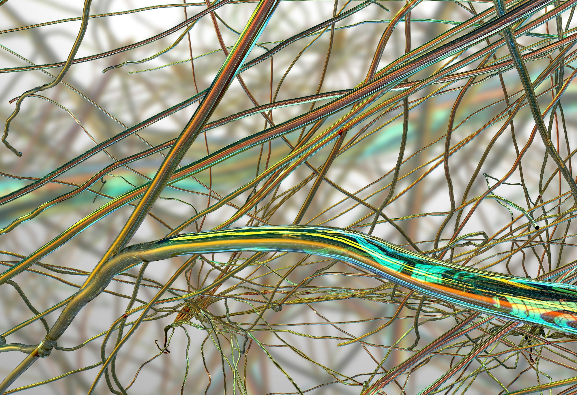
Paper: Photolithographic patterning and mechanical characterization of electrodeposited cellulose nanofiber composite microstructures. Image Credit: Kateryna Kon / Shutterstock
*Important notice: This news reports on an unedited version of an accepted paper and is awaiting final editing. Therefore, the paper should not be regarded as conclusive or treated as established information.
An Article in Press in the journal Scientific Reports demonstrates an electrodeposition-based route for shaping cellulose nanofibers (CNFs) into microscale structures.
The CNF/CMC composite films had an average Young’s modulus of 4.2 GPa, and suspended microbridges were as thin as 300 nm.
The work shows that electrodeposition can be combined with photolithographic patterning to produce submicrometer, free-standing CNF composite structures for further microsystem research.
Bringing Nanocellulose into Microsystems
Cellulose nanofibers (CNFs) are promising bio-based structural materials because they combine low density, high stiffness, mechanical strength, and biodegradability. Researchers obtain these nanoscale fibers by breaking down plant-derived cellulose. These properties make CNFs attractive for microsystem applications, including sensors, actuators, and biomedical devices.
Integrating CNFs into microelectromechanical systems (MEMS) presents several manufacturing challenges. Conventional materials such as silicon support precise microfabrication but remain susceptible to brittle fracture. Metals offer excellent electrical conductivity but can oxidize and fatigue. Polymers offer flexibility, though they may lack sufficient mechanical strength and long-term durability.
CNFs could address some of these limitations, but their integration requires different fabrication strategies. Conventional photolithographic etching can decompose or dissolve cellulose. Their high strength and stiffness also make laser ablation and dry etching difficult.
Previous studies have examined nanocellulose films, hydrogels, and microscale patterning techniques. Few studies have examined thin, free-standing CNF microstructures compatible with standard photolithography.
In this study, the researchers developed an electrodeposition-based approach to fabricate microstructures from mechanically fibrillated CNFs. They introduced carboxymethyl cellulose (CMC) as a co-deposition matrix so that mechanically fibrillated CNFs, not intentionally functionalized with ionizable carboxylate groups, could form cohesive electrodeposited films.
Electrodeposition and Microfabrication Strategy
Mechanically fibrillated CNFs were obtained from kenaf pulp through bleaching and repeated disk milling. TOCNFs were also prepared for comparison. TEMPO-mediated oxidation introduces carboxyl groups onto cellulose fiber surfaces. These groups impart a negative surface charge, allowing TOCNFs to migrate toward the anode under an applied electric field.
The researchers combined an aqueous CNF dispersion with sodium carboxymethyl cellulose. CMC acted as both a dispersant and a co-deposition matrix.
For the thick films used in composition and mechanical measurements, the researchers placed the electrolyte between a copper anode and a platinum cathode and applied a direct-current voltage of 3.0 V for 20 minutes.
During deposition, copper ions released from the anode coordinated with the carboxyl groups in CMC. This interaction promoted network formation, while hydrogen bonding helped incorporate the mechanically fibrillated CNFs into the growing composite film.
AFM and Fourier-transform infrared (FTIR) spectroscopy examined the deposited films' morphology and composition. The CNF fraction was estimated by fitting FTIR spectra.
For microfabrication, the researchers patterned a photoresist mold on a copper-coated silicon substrate. They then electrodeposited the CNF/CMC composite into the patterned regions in two 10-second deposition cycles, with an intermediate 110 °C drying step and a 100 °C, 10-minute drying step before resist removal. A laser Doppler vibrometer measured the fabricated bridges' mechanical resonance under vacuum.
From Composite Films to Suspended Microstructures
CMC enabled the electrodeposition of mechanically fibrillated CNFs. Without the polymeric matrix, the fibers failed to form cohesive films under the experimental conditions. During deposition, the electrolyte produced a blue-green gel-like layer on the copper anode. After 24 hours of ambient drying, water evaporation brought the fibers closer together and promoted hydrogen bonding, producing dense composite films.
Film thickness increased across the prepared electrolyte series as CNF content rose. However, because the CMC mass was held constant while more CNF dispersion was added, total solids concentration also rose, so the experiment cannot separate the effects of CNF fraction and total solids.
AFM observations confirmed that the electrodeposited composite retained its fibrous structure. The CNF/CMC film exhibited a surface roughness of 61 ± 15 nm, compared with 37 ± 10 nm for a drop-cast CNF film. Micrometer-scale aggregates and heterogeneous fiber packing within the CMC matrix contributed to the higher roughness.
FTIR analysis revealed an approximately linear relationship between the CNF concentration in the electrolyte and its fraction in the deposited film.
The researchers achieved a maximum CNF content of 80 wt% using an electrolyte in which CNF accounted for 71.4 wt% of the combined CNF-plus-CMC solids.
The relationship had a coefficient of determination (R²) above 0.95, indicating a close fit to a straight-line trend, and the authors concluded that the CNF fraction in the film could be controlled through the electrolyte composition within the tested range.
Nanoindentation measurements highlighted the reinforcing effect of CNFs. The pure CMC film exhibited a Young's modulus of 2.1 GPa. Incorporating CNFs increased the modulus to 3.9–4.4 GPa across the composite series, averaging about 4.2 GPa. The pure TOCNF film exhibited a comparable modulus of about 4.3 GPa. Increasing the CNF fraction beyond 40 wt% did not further improve stiffness.
The researchers combined photolithography with electroforming to fabricate patterned CNF/CMC structures 300–400 nm thick. The process maintained lateral pattern fidelity even as the material shrank during drying. Thicker deposits also appeared near pattern edges, which the researchers associated with local electric-field concentration and restricted mass transport within the photoresist mold.
A suspended bridge 300 nm thick exhibited a fundamental resonance frequency of 23.34 kHz under vacuum, with a quality factor of 39. This frequency exceeded the calculated stress-free resonance of 7.41 kHz.
Based on resonance modeling, the researchers estimated an effective tensile stress of approximately 172 kPa. They interpreted this inferred stress as likely arising from constrained shrinkage during hot-plate drying before structural release.
The model used a 4.2 GPa modulus from films thicker than 20 µm rather than the 300 nm bridge, and the authors note that differences related to deposition time cannot be ruled out. The 172 kPa value is an effective model estimate, not a direct stress measurement.
Toward Biodegradable Microstructures
The study shows how electrodeposition-based electroforming can integrate plant-derived CNFs into photolithography-compatible microsystems. The team fabricated cohesive films from mechanically fibrillated CNFs while preserving their fibrous morphology.
Mechanical characterization showed that incorporating CNFs reinforces the CMC matrix. The resonance analysis suggests that constrained drying shrinkage likely left residual tensile stress in the fabricated bridges, influencing their resonance behavior.
The authors suggest that CNF-based structures could complement conventional microsystems in uses where environmental sustainability, biocompatibility, disposability, or mechanical damping are priorities.
They suggest applications in disposable biosensors, biodegradable resonant mass sensors, microfluidic mixers, and selected microactuators. The study did not test those applications, nor did it directly measure biodegradation or biocompatibility of the fabricated CNF/CMC structures.
The authors identify environmental stability under humid conditions and thickness control near pattern edges as areas needing further study. The method extends nanocellulose fabrication from conventional films and coatings to precisely patterned, free-standing microstructures. They close by proposing resonators, transient devices, and biodegradable sensing platforms as possible directions for the fabrication method.
Disclaimer: The views expressed here are those of the author expressed in their private capacity and do not necessarily represent the views of AZoM.com Limited T/A AZoNetwork the owner and operator of this website. This disclaimer forms part of the Terms and conditions of use of this website.
Source:
- Kikuchi, K., Van Toan, N., et al. (2026). Photolithographic patterning and mechanical characterization of electrodeposited cellulose nanofiber composite microstructures. Scientific Reports. DOI: 10.1038/s41598-026-77767-0, https://doi.org/10.1038/s41598-026-77767-0