Researchers traced collagen from whole tissue down to nanoscale fibrils to examine how an intact spinal disc responds as mechanical forces move through its complex internal structure.
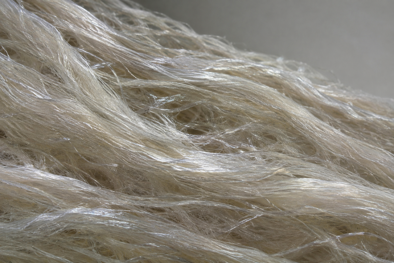
Paper: Multimodal X-ray imaging reveals hierarchical fibre mechanics. AI-generated abstract conceptual image created using ChatGPT/OpenAI
Fibrous composites play an important role in load-bearing structures in advanced engineering materials and biological tissues.
A recent paper published in the journal Nature introduced TomoSAXS, an imaging method that correlates mechanical behavior across multiple length scales within the same intact specimen.
By examining how microscopic collagen fibers deform alongside nanoscale collagen fibril structure under mechanical loading within intact intervertebral discs, researchers mapped aspects of the structural organization and mechanical response of hierarchical materials. The method gives researchers a way to compare nanoscale and microscale mechanics in intact fibrous materials.
Challenges in hierarchical composite analysis
Natural fibrous composites, including tendons, arteries, and spinal discs, combine strength, flexibility, and resilience through hierarchical structures that span several length scales.
These composites organize polymeric molecules and fibers across several structural levels, allowing the resulting structures to withstand repeated, multidirectional mechanical stresses. Studying deformation across these different structural scales remains challenging.
Conventional two-dimensional (2D) X-ray scattering averages diffraction signals across a sample volume, making it difficult to identify directional variations.
Three-dimensional (3D) small-angle X-ray tensor tomography can provide more detailed structural information but typically requires prolonged scanning, sampling around multiple rotation axes, and radiation exposure that can damage sensitive polymer structures.
These limitations make it difficult to directly link nanoscale molecular organization to macroscopic strain and understand how internal stresses are distributed within fibrous materials.
The TomoSAXS methodology: a multimodal approach
Researchers developed TomoSAXS, a multimodal imaging platform that combines phase-contrast computed tomography (pCT), digital volume correlation (DVC), and synchrotron-based small-angle X-ray scattering (SAXS). They conducted experiments at the Diamond Light Source and the European Synchrotron Radiation Facility.
The method uses 3D fiber orientation to separate contributions to overlapping X-ray signals, enabling reconstruction of nanoscale structural information with single-axis sample rotation. Under the chosen conditions, the team reconstructed nanoscale properties for more than 55% of fibers in the scanned volumes.
The experimental workflow began with three-dimensional phase-contrast computed tomography of intact intervertebral disc samples. Automated fiber-tracing algorithms identified individual collagen fiber trajectories and their spatial orientations. The researchers combined these orientations with 3D diffraction models to separate overlapping scattering signals and estimate these properties for individual fibers.
For mechanical testing, the specimens were placed in custom open-frame rigs that maintained hydration with phosphate-buffered saline while applying controlled axial displacements. After a 1-N preload and baseline scans, the researchers applied a 50-µm compressive displacement, allowed at least 20 minutes of stress relaxation, and used DVC algorithms to track displacement, generating detailed maps of local deformation.
To assess the method's applicability beyond rodent samples, the study examined a whole, formalin-fixed human intervertebral disc under static conditions using hierarchical phase-contrast tomography (HiP-CT) combined with high-energy tomographic SAXS.
The human-disc experiment showed that TomoSAXS could reconstruct nanoscale collagen properties across a section of a human-scale specimen, but it did not measure load-induced mechanics in the human disc. The authors state that formalin fixation likely affected the nanoscale collagen properties measured in the specimen.
Pre-strain and molecular mechanics
The multiscale maps linked tissue-level strain, fiber deformation, and fibrillar D-period changes across the disc. Under compression, a 2.2% reduction in disc height produced a mean tissue-level principal compressive strain of 4.2 ± 3.3% in the annulus fibrosus. This result, together with the wide local variation, shows that the tissue deforms unevenly rather than following the bulk strain uniformly.
The study identified an alternating lamellar pre-strain pattern inferred from differences in the approximately 67 nm axial periodicity, or D-period, of collagen fibrils.
Initial D-period, used to infer fibrillar pre-strain, varied inversely with the fibrillar strain produced by loading. Fibrils interpreted as having initial tensile pre-strain shifted toward compression under external loading, while those with initial compressive pre-strain shifted toward tension. The pattern is consistent with pre-strained fibrils moving toward a more neutral state under load.
A separate analysis of the mean and variation in D-period measurements supported a helical-coil model involving molecular loosening or tightening. The study inferred this molecular mechanism rather than observing it directly.
At the microstructural level, fiber strain showed a positive correlation with local structural curvature (r² = 0.480) in the representative disc used for this regression analysis.
The highest curvature occurred mainly in the posterior and lateral transition zone between the central nucleus pulposus and the surrounding annulus fibrosus. The similarity between fiber-level and fibrillar strain magnitudes suggests that much of the deformation was transferred to the fibrils, with limited inter-fibril sliding at the tested loading level.
Assuming a zero-strain D-period of about 67 nm, the paper estimates residual fibrillar strains of about −10% to +10% and pre-stresses of roughly 100 to 200 MPa, comparable with reported failure stresses for isolated rat-tendon collagen fibrils. The authors argue that this mechanically advantageous state may leave less margin before damaging strains are reached.
Biomimetic and synthetic applications
This multiscale imaging method could help researchers test design principles for engineered fibrous materials beyond biological tissues. The paper points to alternating lamellar orientations, localized pre-strain, and strain transfer between structural scales as characteristics that could be examined in synthetic composites.
In aerospace and other composite applications, these principles could help engineers study carbon-fiber composites and nanostructured polymers, including how fiber organization and pre-strain relate to local deformation and damage. Meanwhile, in biomedical engineering, they could help researchers compare tissue-engineered or replacement materials with the structure-function relationships measured in native intervertebral discs, providing a basis for future tests of mechanical compatibility.
TomoSAXS limits and future work
TomoSAXS can examine intact fibrous specimens without physical sectioning while linking nanoscale structure to tissue-scale mechanics. By reconstructing these properties for many traced fibers, researchers could study how aging, fatigue, and stress alter hierarchical systems.
Current scans still require stable samples, and the in situ experiment took about four hours, so the method does not capture the immediate response after loading.
Faster pCT acquisition could help measure time-dependent deformation, while finer angular sampling could increase the fraction of fibers reconstructed.
Radiation exposure also requires further study: the rat in situ protocol delivered a total dose of 80.6 kGy, far below the roughly 2,000 kGy estimated for SAXS tensor tomography with the same setup, but the authors note that the dose may have stiffened collagen fibrils.
Extending the method to nanostructured polymers and carbon-fiber composites would test whether similar multiscale load-transfer patterns occur in engineered systems.
Disclaimer: The views expressed here are those of the author expressed in their private capacity and do not necessarily represent the views of AZoM.com Limited T/A AZoNetwork the owner and operator of this website. This disclaimer forms part of the Terms and conditions of use of this website.