Image Credits: anyaivanova/shutterstock.com
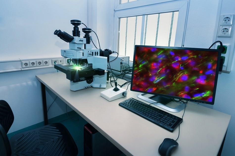
Confocal laser scanning microscopes (CLSMs) use a laser to generate a digital image of a given sample. Confocal microscopes work in tandem with ‘fluorescent tagging’, which involves the alteration of a certain cellular feature to also produce a fluorescent protein of a given color. This allows the observer to visualize specific parts of the sample being studied, and unlike conventional widefield microscopy or electron microscopy better differentiate between different structures within a cell or sample ultrastructure using different colored fluorescent tags.
Fluorescent Tagging
Firstly, common confocal microscopy relies on the use of colored fluorescent proteins which can be encoded into specific genes within an organism or cell. This is carried out through a process known as fluorescent tagging. Building on this, a targeted laser beam is used to excite the fluorescent proteins which would be tagged to the targeted part of the sample under observation. It should be noted that multiple parts of the sample will often be tagged with fluorescent proteins. This means that multiple structures will absorb the photons from the laser beam.
These fluorescent molecules absorb the targeted photon beam - this excitatory beam causes the targeted molecules to fluoresce. The emitted fluorescent light is refracted off of a dichroic (or dichromatic) mirror, followed by several others, which then properly focus this light into a pinhole aperture. This is vital, as it allows only light from the targeted focal plane to be perceived and recorded in the digital display via a photomultiplier tube.
Photomultiplier Tube
The photomultiplier tube, as the name might suggest, perceives the generated fluorescent signal with a photocathode, covered by a protective glass screen. This then amplifies the incoming photon through a collection of anodes and dynodes which bounce the photons along the length of the tube, magnifying the signal, and allowing it to be recorded by the PC program.
Pinhole Optical Screening
The use of the pinhole optical screening technique allows fluorescent emissions which are outside of the focal plane to be selected out and therefore ensures that a high-quality, hugely focused image is generated. This digital rendering is thus free of the visual noise and blurring which is caused by out of focus light which would otherwise also be absorbed.
The downside of this is that in standard confocal microscopy it takes a long time to generate a complete image. Due to the use of only one pinhole, the image must be constructed point by point, pixel by pixel. This means that unlike in standard analog displays, the digital image which is produced takes time to be built up. This problem can be circumvented to a certain degree by utilizing one of two techniques.
Advantages of CLSM
Firstly, some researchers prefer to use a CLSM, which replaces one of the internal mirrors with an Acoustic Optic Deflector (AOD) that controls the beam direction of the fluorescent emission from the sample. This allows more precise targeting than the use of a standard mirror within the microscope chambers.
It is also possible to utilize a CLSM that is adapted for the spinning disk technique. In standard confocal microscopy, only a single pinhole aperture exists to detect the focused beam of fluorescence from the sample. However, spinning disk microscopes use a collection of several thousand pinholes. This allows the image to be rapidly rendered in digital format.
The key to the production of the marvelous 2-D and 3-D images also happens to be one of the greatest advantages of confocal microscopes. Unlike traditional widefield microscopes, CLSMs are ideal for use with thicker specimens, for example, fly embryos and muscle biopsies.
By taking sequential scans of layers of the sample, a complex and highly focused 3-D can be rendered. In addition to this, the application of different colored fluorescent protein tags allows the structure of specific features to be uniquely elucidated. These images can then be placed in relation to each other to produce highly detailed whole-structure 3-D visualizations.
Sources and Further Reading
Disclaimer: The views expressed here are those of the author expressed in their private capacity and do not necessarily represent the views of AZoM.com Limited T/A AZoNetwork the owner and operator of this website. This disclaimer forms part of the Terms and conditions of use of this website.