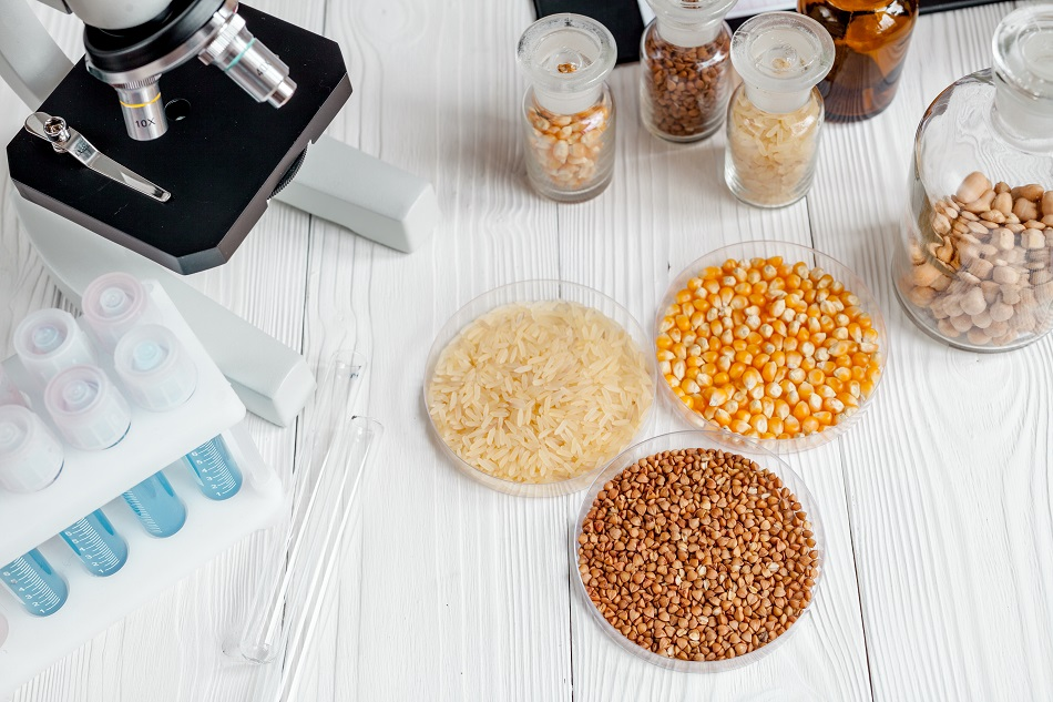
Image Credit: 279photo Studio/Shutterstock.com
Raman spectroscopy (also referred to as Raman imaging) is a useful quantitative tool for analyzing the chemical constituents of a sample. It has been used for a number of years and has gained a notable appreciation among scientists from a different number of fields. Most recently from the nanotechnology sector because of its ability to analyze at the nanoscale.
One area where the use of Raman spectroscopy (including a number of variations) is growing – but is not yet widespread – is in the analysis of foodstuffs because it can analyze and image the microparticles and nanoparticles present within the food sample.
Raman spectroscopy is a method that identifies the vibrational, rotational, and other low-frequency modes of molecules within a sample. These different modes can then be used to determine the chemical constituents of the sample and generate an ‘image’ – or a chemical map of the different molecules within the sample. The vibrational modes are created by shining a beam of light at the sample, the sample scatters the light in a different frequency than the incident light (known as inelastic scattering), and the detection of these wavelengths provides characteristic information of the molecules (and their bonding) within the food sample.
One key feature of Raman imaging methods that makes them a highly useful class of analysis methods for the food industry is that they are not sensitive to high water contents. Almost all foodstuffs have some water content (with some foodstuffs having a lot) and this can affect the sensitivity and operational effectiveness of some analysis methods. The analyses possible with Raman imaging make it an ideal method for analyzing one-off samples, as well as being used on food processing lines as a quality control (and monitoring) tool.
Raman imaging can be used to determine: a wide range of micron-sized and nano-sized particulate matter within foodstuffs (including additives); the particle size and chemical structure of the foodstuffs at these dimensions; and to check that the packaging material has not leaked any particulate matter into the food. Overall, it is a method that can be used with a wide variety of solid, liquid, and complex mixture foodstuffs.
One of the biggest areas of use is to check for food contamination, which can come from the packaging as well as at the agricultural stages of the food product’s life cycle. The foodstuffs at the original agricultural source can also be checked to see if any pesticides, harmful substances, or bacteria are present in the foodstuff (and this can be done in the processing stages as well either in a continuous manner or in individual batch checks).
Outside the manufacturing environment, quality control tests at land and sea borders use Raman imaging methods to detect whether any of the imports/exports contain substances that are prohibited in the intended product destination. Because this is done on a few individual products (and not on all the products), the specific tests use dyes, or other binding substances, which will bind to any of the specific contaminants that are prohibited and provide a clear analysis as to whether those molecules are present in the food product.
Confocal Raman Spectroscopy
Confocal Raman spectroscopy is more widely used when a more accurate representation of the chemicals structures in the food sample is needed, that is to say, it is a highly magnified and more sensitive version of conventional Raman spectroscopy. As well as being used on specific foodstuffs, such as bananas, chocolates, and candies, confocal Raman spectroscopy is widely used to analyze the additives which are added into food. Commonly, these include emulsifiers, stabilizers, carbohydrates, or thickeners in foodstuffs such as those found in instant gravy and dairy emulsions.
It’s a technique that’s often widely used to identify and image the phase separation of different foodstuffs, to generate images of the surface topography of sugary foods, and to give a 3D representation of the chemical species distribution throughout a food sample.
Surface-Enhanced Raman Spectroscopy (SERS)
SERS is a more sensitive version of Raman spectroscopy that utilizes the excitation of localized surface plasmons to increase the sensitivity of the measurement. This extra sensitivity enables SERS to analyze and identify molecules in trace amounts (rather than in the larger concentrations seen with other Raman imaging methods). For foodstuffs, either silver or gold nanoparticles are used to improve the sensitivity (as the surface plasmons) so that the organic molecules in the food can be imaged at the parts per million (ppm) level.
Overall, the application of SERS to foodstuffs includes the detection of pesticides present in fresh food (such as fruits); the determination of fat/oil composition in a food sample; whether oil has been adulterated; whether a foodstuff contains any harmful bacteria (or another micro-organism); for detecting carotenoids; for detecting the levels of melamine in milk; and for analyzing the structure of grains and crops, and more.
Sources and Further Reading
Disclaimer: The views expressed here are those of the author expressed in their private capacity and do not necessarily represent the views of AZoM.com Limited T/A AZoNetwork the owner and operator of this website. This disclaimer forms part of the Terms and conditions of use of this website.