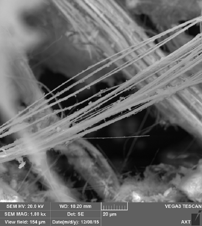
Asbestos samples imaged by Dr. Kamran Khajehpour from AXT Pty. Ltd. Using a TESCAN VEGA3 SEM
The health risks caused by damaged asbestos are so high that it has a disease, asbestosis, named after it. Asbestosis is a type of pulmonary fibrosis, where high levels of exposure to asbestos fibers lead to extensive scarring of the lungs.
Mild cases may result in wheezing and shortness of breath, but more severe complications include heart diseases and mesothelioma, a cancer of the tissue layers that cover the internal organs.
Despite UK legislation banning the sale and use of white asbestos in 1999 with few exemptions, asbestosis still claims the lives of over 5000 workers a year. Given that it typically takes 20 – 30 years post-exposure for asbestosis to develop, it is likely that the benefits of the 1999 legislation will only start to lead to a reduction in deaths now.
Evaluation of whether legislative and workplace protection methods have been successful relies on unambiguous identification and diagnosis of asbestosis. This is also essential for litigation cases of injured workers to be successful. Traditionally, a combination of chest radiographs with an occupational history of working with asbestos would have been considered sufficient for a diagnosis of asbestosis. However, there have been suggestions that chest radiography does not give a truly unambiguous diagnosis, particularly in the early stages of the disease. Alternative diagnostic techniques include highly invasive chest biopsies and analysis of the asbestos fibers using light and transmission microscopy techniques.
How can Electron Microscopy Identify Asbestos in Patients?
Recent work by Dr. Takashi Kido and co-workers has shown that electron microscopy may be a powerful tool for identifying whether asbestos fibers and particles are present in patients with diffuse lung diseases, making it possible to conclude whether the lung disease was likely asbestos-exposure related.
As well as this, electron microscopy made it possible to detect and identify the presence of elements such as iron and phosphorous. These elements can be used as markers of the levels of occupational exposure a patient had undergone, enabling future studies examining the relationship between levels of exposure and negative health consequences.
Asbestos Fibers Under the Electron Microscope
The identification of asbestos fibers and particles is often carried out on lung biopsy samples. This is because the tissue typically contains large amounts of fibers, making detection and identification more straightforward. Fibers generally have a distribution of sizes and shapes, but careful analysis of the fiber type and morphology can be useful in the identification of asbestos sources or differentiation between exposure to different types of asbestos.
However, lung biopsies are an invasive medical procedure, carrying a risk of pneumonia and lung collapse in patients who normally already have impaired lung function. Being able to retrieve the same information using bronchoalveolar lavage fluid, which is obtained by washing the lungs with a small volume of fluid via a bronchoscope, therefore reduces patient risk and discomfort.
Dr Kido and the team found that electron microscopy studies on bronchoalveolar lavage fluid allowed for much more sensitive detection of asbestos than light microscopy. This improvement in sensitivity meant that using electron microscopy on the dilute fluid samples was as effective as using light microscopy on lung biopsy samples that had much higher asbestos concentrations.
Electron Beams in Microscopies
Electron microscopy uses electron beams, rather than photons as in the case of light microscopy, to image a sample. One of the advantages of electron microscopy is it typically offers much greater levels of magnification, making it possible to resolve smaller objects and can have very high image resolution so more details on the shape and size of objects can be revealed.
Click here to find out more about electron microscopy equipment
The images recorded on the electron microscope can be used to identify the number of fibers in the sample, and therefore the asbestos concentration, their size, but also their elemental composition, which is very challenging to do with light-based techniques. This is because the incident electron beam is composed of very high energy electrons, some of which will be absorbed by the sample.
When the incident electrons are absorbed, their energy is transferred to the electrons in the atoms of the sample. This causes the atomic electrons to be excited, or even ejected, leaving the atom in an unstable state. This means it becomes energetically favorable for an electron to collapse and fill the vacancy left by the previously excited electron. However, to conserve the overall energy, this results in the emission of another electron or even a photon.
The emitted radiation occurs at energies that are characteristic of specific elements and so can be used to identify the elemental composition of the sample, not just the shape and size of the particle or fiber. By using the elemental information against exposure history, Dr. Kido and colleagues could show that electron microscopy was a potential tool for identifying previous exposure levels.
By demonstrating that electron microscopy could be used to detect asbestos fibers from bronchoalveolar lavage fluid as light microscopy on lung biopsies suggests that, with a change of analysis technique, electron microscopy fluid analysis may have the potential to replace lung biopsies as the gold standard for asbestos exposure analysis. The elemental-identification power of electron microscopy will also open the possibility of the identification of the role of particular minerals in asbestos-related lung diseases.
What is transmission electron microscopy and its advantages? Click here to find out more.
Sources and Further Reading
- 1999 Asbestos Regulations, http://www.legislation.gov.uk/uksi/1999/2373/made, (accessed 17/03/2020)
- Asbestosis Related Disease Status 2019, https://www.hse.gov.uk/statistics/causdis/asbestos-related-disease.pdf, (accessed 17/03/2020)
- Attanoos, R. L., Alchami, F. S., Pooley, F. D., & Gibbs, A. R. (2016). Usual interstitial pneumonia in asbestos-exposed cohorts – concurrent idiopathic pulmonary fibrosis or atypical asbestosis? Histopathology, 69(3), 492–498. https://doi.org/10.1111/his.12951
- Ross, R. M. (2003). The clinical diagnosis of asbestosis in this century requires more than a chest radiograph. Chest, 124(3), 1120–1128. https://doi.org/10.1378/chest.124.3.1120
- Roggli, V. L. et al. (2010). Pathology of asbestosis an update of the diagnosis criteria.pdf. Arch Pathol Lab Med, 134, 462.
- Kido, T., Morimoto, Y., Yatera, K., Ishimoto, H., Ogoshi, T., Oda, K., … Mukae, H. (2017). The utility of electron microscopy in detecting asbestos fibers and particles in BALF in diffuse lung diseases. BMC Pulmonary Medicine, 17(1), 1–9. https://doi.org/10.1186/s12890-017-0415-5
Disclaimer: The views expressed here are those of the author expressed in their private capacity and do not necessarily represent the views of AZoM.com Limited T/A AZoNetwork the owner and operator of this website. This disclaimer forms part of the Terms and conditions of use of this website.