In archaeological and paleontological research, unique mineral artifacts, such as stone tools, bone specimens, and fossils often require specialized non-invasive yet detailed analysis. Atomic force microscopy (AFM) is one of the analytical methods that can provide a non-invasive, non-destructive, and nanometer-scale characterization of archaeological and paleontological samples.
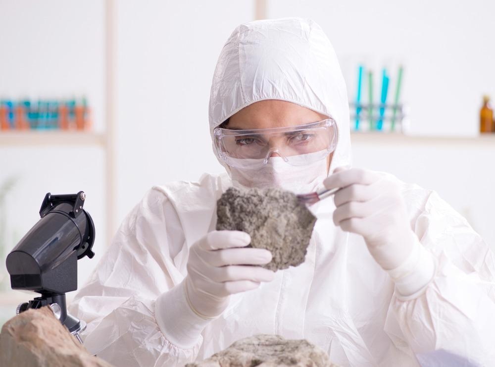
Image Credit: Elnur/Shutterstock.com
The development of AFM in the late 1980s gave scientists a powerful high-resolution analytical technique that rapidly became a mainstay method in material science, the semiconductor industry, life sciences, and many other research fields.
Non-Invasive Analysis at the Nanoscale
The technique is non-invasive and non-destructive and enables the measurement of the surface topography of a sample by tracing the sample surface with an ultra-sharp probe attached to a soft spring-like cantilever. When the probe approaches the sample surface, weak interaction forces, such as Van der Waals and electrostatic forces, occur between the probe and the sample.
The resulting perturbation in the cantilever motion is recorded and converted into a three-dimensional representation of the sample surface. In addition, AFM can detect the response of the probe to the viscoelastic properties of the sample.
One main advantage of AFM is the ability to operate and provide three-dimensional information with sub-nanometre spatial resolution under ambient conditions (in air or liquid) without any staining or other sample modifications. This is particularly beneficial when analyzing biological or other equally fragile and delicate specimens.
Microwear Traces Elucidate the Way Our Predecessors Made and Used Tools
In the past two decades, the non-invasive nature of the AFM technique attracted interest from the archeological research community. In archaeological research, AFM can provide quantitative information when studying the microwear polishes of archeological artifacts at a sub-micron scale.
Importantly, unlike other micro and nanoscale techniques, such as scanning electron microscopy (SEM), AFM can provide quantitative roughness analysis without sputter coating or casting a replica of the delicate artifact. The AFM enabled the scientists to obtain images with higher spatial resolution compared to optical interferometry or laser scanning confocal microscopy, techniques commonly used for quantitative analysis of stone tools and other mineralized artifacts.
Several research groups successfully employed AFM for quantitative microwear analysis of Paleolithic stone tools and flints. By quantitative studies of the surface roughness and morphology on the tool surface, the researchers were able to conclude that depending on the use of the tool (whether it was used to cut meat, wood, bone, or others) the microwear polish can exhibit very distinct morphology and surface roughness when compared to an unused control region on the tool. Such information is essential when evaluating the possible use of the tools and the evolution of their development.
Nanoscale Characterization of Plant and Unicellular Fossils
Sub-micron scale characterization of fossils demonstrated the versatility of AFM for paleontological research.
The technique enabled an international team of researchers from Ludwig-Maximilians-Universität in Munich, Germany, and the University of Alabama and UCLA, in the United States, to analyze the fine structure of organic-walled Precambrian fossils, such as microscopic sphaeromorph acritarchs (small organic-walled microfossils) and planktonic unicellular protists cysts (dormant stage of microscopic unicellular organisms) permineralized around 650 million years ago in the Chichkan formation in southern Kazakhstan.
Detailed AFM observation revealed that the fossil walls consisted of stacked arrays of 200 nm-sized angular platelets of polycyclic aromatic kerogen. In another study, the researchers compared a fossil cell wall nanostructure obtained by AFM with data obtained by SEM of identical samples.
The results demonstrated that, while the SEM and AFM displayed similar features, the AFM offered higher resolution and three-dimensional information about the organization of the kerogen in the cell wall, consisting of multi-lamellar amorphous carbon films composed of layers with thickness between 10 nm and 20 nm.
Image Credit: Vigni Michael/Shutterstock.com
With the help of the AFM technique, several other research groups identified crystal structures of biogenic marine calcite and aragonite in fossil corals, shells, and algae. The AFM provided detailed information on the nanotexture, distribution, orientation, and shape of these biominerals.
The technique has been also used to characterize fossil specimens of higher vascular plants, such as fossil cytoplasm of charcoalified coniferous cones. For the first time, the scientists were able to observe fossilized subcellular features such as a nucleus-like region, small granules in the cytoplasm, and different organelles with membranes that corresponded with those of live plant cells.
Direct Imaging of Library Materials Degradation
A research team from the University of Rome demonstrated that the AFM technique could be indispensable when evaluating the conservation state of paper and paper-like artifacts. The scientists were able to distinguish paper where the surface cellulose fibers were well-preserved from paper that already underwent some degradation process.
Such subtle differences are usually hard to measure with other analytical techniques or require the application of destructive techniques to be detected. Further, it was possible to identify the presence of materials other than cellulose fibers and fibrils, such as dust and microorganisms, thus allowing identification of the processes affecting the preservation of the paper specimens. Such applications highlight the potential of AFM for early detection of library material deterioration and better preservation of cultural heritage research.
The growing adoption of AFM in the field, especially when combined with other microscopic (optical, SEM) and spectroscopic techniques (Raman spectroscopy), can make a valuable contribution towards a better understanding of the history and evolution of archaeological and paleontological samples, and the geochemical maturation and preservation of organic matter based on the sample surface morphology and physical properties.
References and Further Reading
Faulks, N.R., et al. (2011), Atomic force microscopy of microwear traces on Mousterian tools from Myshtylagty Lagat (Weasel Cave), Russia. Scanning, 33, 304-315. Available at: https://onlinelibrary.wiley.com/doi/epdf/10.1002/sca.20273
Piantanida, G., et al. (2005) Atomic force microscopy imaging directly on paper: a study of library materials degradation. Proc. SPIE 5857, Optical Methods for Arts and Archaeology, 58570R. Available at: https://www.spiedigitallibrary.org/conference-proceedings-of-spie/5857/1/Atomic-force-microscopy-imaging-directly-on-paper--a-study/10.1117/12.612099.short?SSO=1
KKempe, A., et al. (2002) Atomic force microscopy of Precambrian microscopic fossils. Proc. Nat. Acad. Sci. USA, 99 (14), 9117-9120. Available at: https://www.pnas.org/content/99/14/9117
Disclaimer: The views expressed here are those of the author expressed in their private capacity and do not necessarily represent the views of AZoM.com Limited T/A AZoNetwork the owner and operator of this website. This disclaimer forms part of the Terms and conditions of use of this website.