A thorough comprehension of cellular bio-functionality necessitates knowledge about specific constituents of the cell membrane and, as a result, the structure and organization of protein complexes, as well as tools for high-resolution investigations. Such high-resolution research studies are possible via the utilization of the freeze-fracture technique's accurate portrayal of the membrane via breakage and replication.
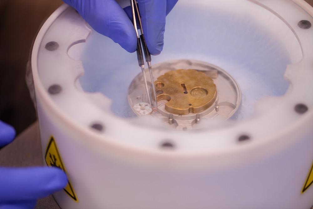
Image Credit: PolakPhoto/Shutterstock.com
Brief Introduction and Methodology
The method of fracturing a frozen material to disclose interior features is known as freeze-fracture. It entails physically separating (fractioning) a frozen biological sample and disclosing structural information via the fracture surfaces using transmission electron microscopy. Biological membranes possess an inherent plane of fragility in their hydrophobic core when frozen, therefore if the specimen is shattered or fractured, the fracture region will frequently separate the barrier into half-membrane lobes.
The outcome is a three-dimensional image of the cell's plasma membrane arrangement, complete with perspectives of the barrier inside. By creating a very precise platinum-carbon duplicate of the fracture plane, these features may be seen under the electron microscope.
Historical Origins and Development
For more than 50 years, freeze-fracture electron microscopy has been widely accepted as a significant method in microscopic structural and biological research. Although components of the approach were present in its early stages as early as the 1950s, its potential attracted the attention of cell biologists around 1960.
Originally, explanatory conflicts hampered the use of freeze-fracture, but once these were remedied, the method prospered during the 1970s and 1980s, offering breakthroughs in the interpretation of the structural organization of cellular membranes and plastids that traditional thin-section electron microscopy could not achieve.
Steps
There are four processes to creating a freeze-fracture replica. The specimen must be rapidly frozen in the initial stage. The specimen is then aggressively fractured at low temperatures. The third phase is vacuum coating of platinum and carbon to create a duplicate of the frozen cracked surface, followed by washing the replica to eliminate any biological material. Furthermore, the freezing process is frequently performed after pretreatment, and a supplementary etching step may be included between fracturing and replica creation.
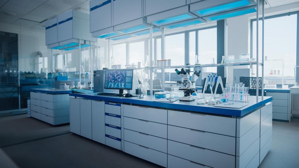
Image Credit: Gorodenkoff/Shutterstock.com
The usual approach for fast freezing is to quickly submerge a properly affixed specimen in a liquid coolant (e.g., subcooled liquid nitrogen). Regrettably, until they are pretreated previously with a cryo-preservative, the majority of the biological samples preserved in this manner display ultrastructural ice crystal degradation. Glycerol is the most often used cryoprotectant.
The replica is created in two steps: shadowing and backing. In the usual approach, oblique, directional shadowing is accomplished by vaporizing a thin coating of platinum–carbon onto the sample, swiftly followed by an electron-lucent carbon reinforcing (backing) film condensed from above. The geomorphological elements of the frozen, broken surface are thereby transformed into changes in the depth of the replica's implanted platinum covering. After the replica is obtained, the specimen is exposed to standard atmospheric pressure and subjected to ambient temperature. Sodium hypochlorite solution, chromic acid, or other cleaning chemicals are used to eradicate biological molecules from the replica.
Advantages
Freeze-fracture electron microscopy (FEM) is a one-of-a-kind research technology that offers a slew of significant advantages to microbiologists. For starters, because it is a duplicating method, it may offer both face-on and cross-sectional perspectives of biological components, letting the researchers precisely observe the architecture and dispersion of particular features both inside the fracture plane and on the membrane surface.
Even when biochemical preprocessing is utilized, it gives an additional technique of further processing, allowing for the comparison of thin-section electron microscopy and FEM statistics. Furthermore, when combined with a mere physical sample preservation approach (such as cryofixation), FEM can fully eradicate the necessity for biochemical fixation, making it particularly useful for the detection of artifactual features.
Methods and Uses
For the processing of biomaterial, two distinct freeze-fracture procedures have been investigated. In the first method, biological tissue is first fixated and dried in absolute alcohol, then frozen, fragmented, defrosted in absolute alcohol, and critical-point dried. This approach produces great results when examining the lumen region of cells that border natural anatomical gaps such as the liver sinusoids or Bowman's capsule in the kidney. This initial approach, in general, revealed little information about the cytoplasmic membrane or nucleolus of cells, and thus a second method was explored.
The second method involves tissue samples or lymphocytes being initially injected with cryo-preservative, frozen, shattered, and not repaired until after melting. Finally, the fixed cells are dehydrated. This particular approach has been utilized to investigate mammal tissues, carrot protoplasts, and leaf meristematic cells.
Equipment for Freeze Fracture Electron Microscopy
The Freeze Fracture System Leica EM ACE900 is effective equipment for this process. The Leica EM ACE900 sample preparation system can carry out freeze fracturing, freeze etching, and e-beam coating without the use of any extra tools.
What is Ultrarapid Freezing?
Although several types of specimens with low moisture concentration can be effectively preserved by normal plunge freezing without preceding cryoprotective processing, rapid freezing in most cases necessitates ultrarapid freezing procedures. These procedures are intended to increase the cooling rate of the specimen substantially to prevent the ice crystal deterioration that occurs in non-pretreated samples. Optimal plunge freezing, jet freezing, and spray freezing are the main ultrarapid freezing procedures.
Latest Research
The most recent study in the journal Star Protocols describes how to use FEM to locate Phosphatidylcholine (PtdCho), which is freshly generated via the Kennedy route in yeast. The endoplasmic reticulum synthesizes PtdCho, a key transmembrane phospholipid. The methodology includes administering a clickable alkyne-containing choline analog to lymphocytes, quick-freezing, freeze-fracture replica production, conjugation, and immunogold labeling. This approach may be used to quantify which barrier flaps have freshly generated PtdCho integrated.
In recent times, new methods of freeze-fracture microscopy techniques are being studied and developed, such as the high-pressure freezing technique. These methods will overcome the traditional limitations, paving the way for researchers related to the fields of genetics, bioengineering, and the medical industry.
More from AZoM: What is Coordinate Metrology?
References and Further Reading
Tsuji, Takuma, and Toyoshi Fujimoto. 2021. Ultrastructural localization of de novo synthesized phosphatidylcholine in yeast cells by freeze-fracture electron microscopy. STAR protocols. 2(4). 100990. Available at: https://www.sciencedirect.com/science/article/pii/S2666166721006961?via%3Dihub
Leica Microsystems, 2022. Freeze Fracture System Leica EM ACE900. [Online]
Available at: https://www.leica-microsystems.com/products/sample-preparation-for-electron-microscopy/p/leica-em-ace900/
Meier, Carola, and Anja Beckmann. 2018. Freeze fracture: new avenues for the ultrastructural analysis of cells in vitro. Histochemistry and Cell Biology 149(1).3-13. Available at: https://link.springer.com/article/10.1007/s00418-017-1617-x
Kaech, Andres, and Urs Ziegler. 2014. High-pressure freezing: current state and future prospects. Electron Microscopy. Humana Press, Totowa, NJ. 151-171. Available at: https://link.springer.com/protocol/10.1007/978-1-62703-776-1_8
Severs, Nicholas J. 2007. Freeze-fracture electron microscopy. Nature protocols 2(3). 547-576. Available at: https://www.nature.com/articles/nprot.2007.55
Disclaimer: The views expressed here are those of the author expressed in their private capacity and do not necessarily represent the views of AZoM.com Limited T/A AZoNetwork the owner and operator of this website. This disclaimer forms part of the Terms and conditions of use of this website.