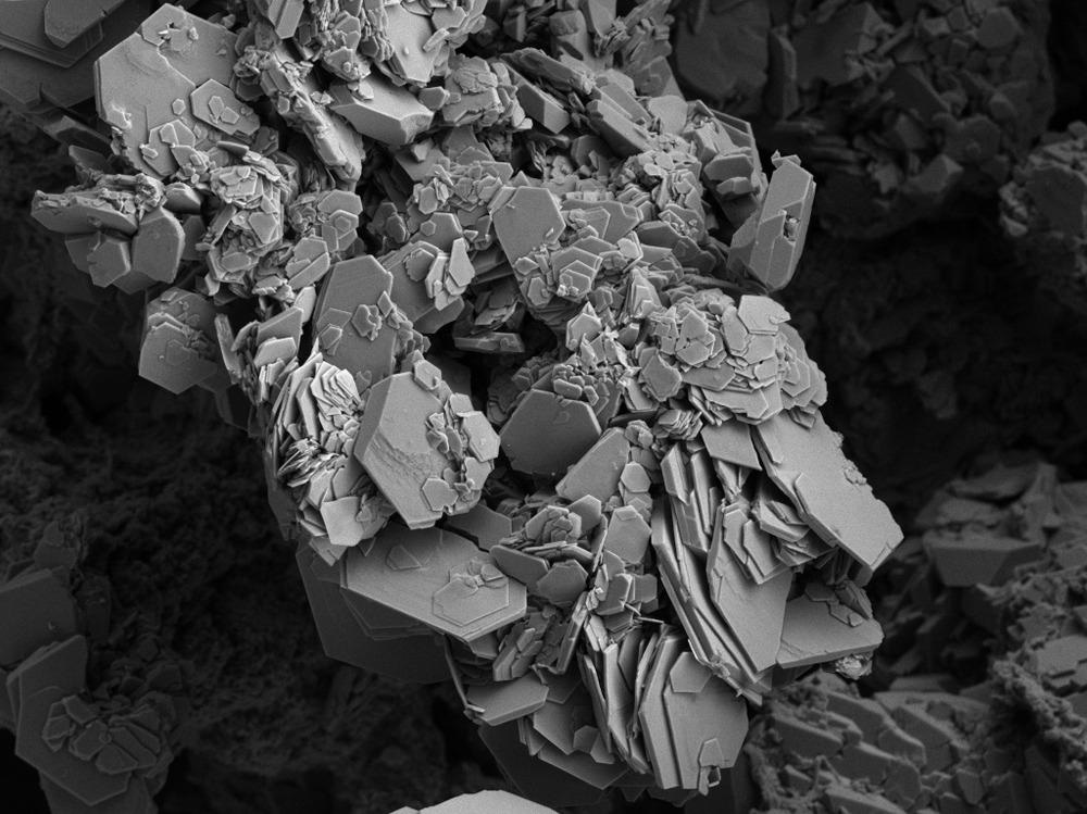
This article focuses on the use of electron microscopy paired with fast Fourier transform (FFT-EM), its advantages and limitations, alternative methods, and recent studies involving this method.

Image Credit: Ravenash/Shutterstock.com
What is FFT-EM?
FFT-EM is an innovative method that represents a combination of FFT and EM techniques such as scanning electron microscopy (SEM). The technique is often used to determine the interior and surface planar pore structures, defect structures, and plane groups of organized pore arrangements in several mesoporous materials. FFT-EM can provide a simple and unique approach to characterize both defects and pore structures of bulky mesoporous materials with different complex structures.
Mesoporosity is the most crucial factor in determining the effectiveness of mesoporous materials for different applications such as bio-imaging, nanosynthesis, and drug delivery. Mesoporosity is referred to as the three-dimensional (3D) or interior and surface cross-sectional structure of the pores. The pore order must be determined to identify new mesoporous materials or thoroughly understand the self-assembly mechanism during the mesoporous material syntheses.
Alternative Methods to FFT-EM
High-resolution scanning electron microscopy (HR-SEM) and transmission electron microscopy (HR-TEM) can be used as alternatives to FFT-EM. Although HR-SEM can be used for producing two-dimensional (2D) projections and surface imaging, HR-TEM imaging is a more suitable technique to directly observe a volume of atoms. Additionally, TEM can provide an effective way to visualize the interior pore interconnectivity. Thus, the HR-TEM technique, typically with small-angle diffraction (SAD), is used extensively in studies that investigate the plane group and structure of pores in mesoporous materials.
Although TEM is a widely used and powerful technique, it has certain disadvantages. For instance, the images obtained using TEM only provide 2D information on the pore structure. 3D orientation of mesopores can be obtained only through a complex program where a series of electron diffraction (ED) patterns and HR-TEM images of the same position at various tilt angles are collected and then a computational image reconstruction is performed.
The quality of images obtained through the HR-TEM technique can be influenced by the equipment condition and sample preparation method. Moreover, the high voltage used in HR-TEM imaging can quickly damage the ordered pore structure of the mesoporous material to a significant extent, which can affect the entire imaging process.
Low-voltage SEM can be used to obtain sub-nanometer scale resolution, which provides a new way to investigate the mesoporous material pore structure. Currently, SEM is primarily used to measure the pore/particle size and determine the morphological features and phase composition on the material surface. However, the technique can also reveal the arrangement of sub-surface or surface pores. Thus, the technique is often used with TEM to determine pore structure.
Additionally, the electron backscattered diffraction (EBSD) of SEM can provide structural and compositional information at an atomic level as it can determine the interplanar angles and spacing of crystallized materials. However, the ordered mesopore arrangement in silica materials is intrinsically different from the atomic level ordering in conventional crystals. Thus, EBSD cannot determine the space groups of the ordered pore arrangements.
Advantages of FFT-EM Technique over TEM and SEM
The FFT-EM strategy can effectively characterize the pore structure and inner defects. In this technique, the pore arrangement obtained in the cross-sectional SEM images is determined by FFT analysis, while the cross-section ion milling is used to characterize the interior defects. The technique also allows the combination of several interior planar images of similar orientations to achieve a 3D reconstruction of the pore structures.
A low electron beam current and acceleration voltage can be used during the HR-SEM to minimize the damage to the mesoporous materials due to the electron beam. Image J, a Java image processing program, is used to process the SEM images through FFT analysis to obtain a Fourier diffractogram. The FFT analysis transforms the greyscale distribution function of the SEM image into a frequency distribution function.
A diffraction pattern can be obtained from the frequency distribution function, which can be used to effectively determine the symmetry and periodicity of the pore structures. The pore crystal lattice plane can also be determined by the FFT analysis of SEM images collected through constant tilting of the mesoporous material sample until the live FFT matches the pore crystal structure pattern.
Limitations of the FFT-EM Technique
FFT can transform a limited range of waveform data, and a window weighting function has to be applied to the waveform to compensate for the spectral leakage, which can reduce the overall effectiveness of the FFT-EM technique.
Recent Studies Involving this Technique
In a study published in the journal Microporous and Mesoporous Materials, researchers used the SEM technique paired with the ion milling process and FFT to successfully characterize the interior defects and mesopore structure ordering of large pore silica nanospheres, KIT-6, and SBA-15 mesoporous silica samples. The FFT-SEM technique demonstrated different pore arrangements in the subsurface, surface, and interior of mesoporous materials and determined the plane groups of crystals.
To summarize, EM paired with FFT has emerged as one of the effective techniques for the determination and reconstruction of pore structures. However, more research is required to eliminate the limitations associated with this FFT-EM to use the technique in more applications.
More from AZoM: What Multi-Analytical Techniques are Used to Assess Paintings?
References and Further Reading
Xu, F., Wu, W., Liu, Z. et al. Combining scanning electron microscopy and fast Fourier transform for characterizing mesopore and defect structures in mesoporous materials. Microporous and Mesoporous Materials. 2015.
Yi Zeng, Ziwei Liu, Wei Wu, Fangfang Xu, Jianlin Shi. Combining scanning electron microscopy and fast Fourier transform for characterizing mesopore and defect structures in mesoporous materials. Microporous and Mesoporous Materials. Volume 220, 2016, Pages 163-167. ISSN 1387-1811. https://doi.org/10.1016/j.micromeso.2015.09.001
What is the main disadvantage of FFT? [Online].
Disclaimer: The views expressed here are those of the author expressed in their private capacity and do not necessarily represent the views of AZoM.com Limited T/A AZoNetwork the owner and operator of this website. This disclaimer forms part of the Terms and conditions of use of this website.