A research group at Uppsala University, Sweden has developed a new responsive coating for implants used in surgery to improve their integration into bone and to prevent rejection. Neutron scattering experiments at the Institut Laue-Langevin (ILL) in Grenoble, France have shown how a protein that promotes bone growth binds to this surface and can be released in a controlled way.
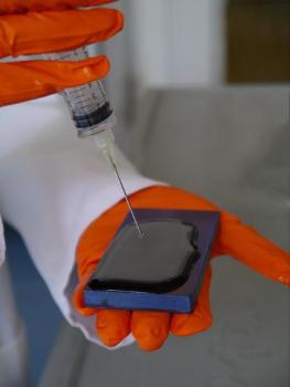 A gel coated titanium surface binds proteins which promote bone formation. Credit: Ida Berts
Orthopaedic and dental implants must last for many years. Success for these surgical components depends on integration into adjacent bone tissue. Gels made by modifying hyaluronan, a large biological molecule, can be used to coat implants. A new paper in Advanced Engineering Materials shows that the coated titanium surfaces can bind protein molecules which promote bone formation. These can be released slowly once the surface comes in contact with a solution of calcium ions. This process would stimulate the growth of bone on the implant.
The gel layers, a few millionths of a millimetre thick, were characterised using neutron reflection at the ILL, a technique that provides a detailed picture of what happens at a surface. In their new paper the research team showed that the protein, BMP-2, that encourages bone growth was bound to the gel. They also demonstrated that the layer of protein was stable in water but could be released slowly by adding solutions containing calcium, a process that was observed in real time using neutron reflection to track the amount of protein at the surface.
The research group has now launched trials of similar materials for metal implants in rabbits. These ongoing studies are made in collaboration with the Swedish Agricultural University in Uppsala and they provide a step towards transfer of the results to clinical applications.
'Interdisciplinary research and partnerships allow advanced analytical tools to be applied to important but difficult medical and scientific challenges. This exciting work comes from shared goals of chemists and physicists as well as the Centre for Neutron Scattering at Uppsala University and the laboratories in Grenoble', says Professor Adrian Rennie.
'We envisage that the materials will be used in medicine to modulate the healing process in bone', says Associate Professor Dmitri Ossipov. He continues, 'Neutrons are an ideal tool to understand the interactions of metal surfaces, polysaccharide biopolymers, and proteins thanks to a contrast matching technique that highlights only the protein components at the interface.'
'Neutron scattering techniques are increasingly relevant to optimise bio-materials and to study systems that relate to health. The importance of combining conventional laboratory studies with those at a large scale facility to give a complete picture of a process was proven once more. This work arose from a studentship funded by the Institut Laue-Langevin which makes us proud of our PhD programme.' says Dr Giovanna Fragneto from the Institut Laue-Langevin.