Cardiovascular disease is one of the leading global causes of death, placing a huge burden on healthcare systems worldwide. In 2017, around 18 million people worldwide died from cardiovascular disease. In the US alone, according to the Centers for Disease Control, someone has a heart attack every 40 seconds, with about 805 thousand people suffering from a heart attack annually.
Types of cardiovascular disease include ischemic heart disease and congenital heart disease lead to a lack of cardiac function, and adult myocardium does not have the ability to heal itself after injury. There are several strategies currently to treat heart disease, including transplantation in the most severe cases. Common treatments for myocardial infarction only prolong life and relieve symptoms rather than restore damaged and lost myocardial tissue.
Additionally, transplantation can be problematic. There is a risk that the patient will reject the donor organ as well as there being a risk of complications during and after heart surgery. Moreover, there is a critical lack of donor organs, leading to waiting times for patients and increasing the risk of fatalities. Current treatments are lacking in effectiveness, and long-term regeneration of heart tissue, which is an eminently preferable medical outcome, is currently not possible in practice.
Stem Cells and Regenerative Medicine
Innovative approaches have been explored in recent years to achieve the regeneration of damaged or lost tissue, leading to the development of the field of regenerative medicine. Stem cell therapies are at the heart of the field. Several studies have demonstrated that stem cell therapies have advantages for regenerating heart tissue, including enhancing cardiac function, reducing infract size, and improving angiogenesis.
However, even this field has its limitations when it comes to the long-term restoration of cardiac tissue. Stem cell therapies are hampered by limitations intrinsic during post-transplantation, such as low cell retention, the low survival rate of engrafted cells, and a lack of host tissue regeneration. For this reason, in vitro approaches to applying cutting-edge technologies to construct functional cardiac tissue are a current focus in tissue engineering research.
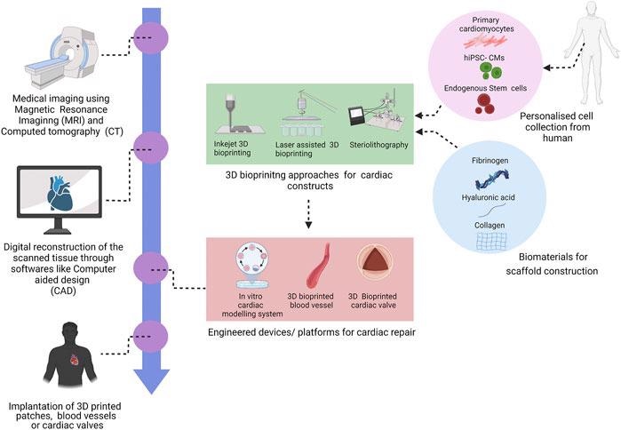
3D bioprinting technology for cardiac repair and regeneration. Image Credit: Chingale, M, et al., Frontiers in Materials
The Study: 3D Printing Functional Cardiac Tissue
The new study published in Frontiers in Materials has explored the use of 3D bioprinting to manufacture functional cardiac tissue. This innovative approach to tissue engineering has the potential to significantly improve the quality of life and life expectancy for patients suffering from cardiovascular disease worldwide. The study provides an overview of 3D bioprinting techniques, potential applications, limitations, and future prospects.
The field of cardiovascular tissue engineering brings together cell biology, material science, and biofabrication to achieve regenerative therapeutics. Bioprinting both two- and three-dimensional tissues and structures utilize live cells, biomaterials, and bioactive molecules for in vivo and in vitro applications. 3D printing can construct biomimetic structures which possess the intricate cellular composition and matrix structure of cardiac tissue.
Recent studies in the field of tissue engineering have used 3D bioprinting techniques for multiple research applications, including regenerative studies and drug screening. Additionally, 3D bioprinted aortic valves and conduits have been used in bypass procedures and the treatment of aortic valve diseases. 3D cardiac patches can preserve the functionality of cardiac tissue after ischemic events.
Further research in the field has explored the use of 3D printed vascular grafts to enhance biocompatibility and structural stability. Another study has demonstrated that a 3D bioprinted cardiac patch constructed from human coronary artery endothelial cells, with methacrylate micropatterning and an alginate matrix, possesses high cell proliferation, migration, and differentiation. The field of cardiac tissue engineering has some interesting research directions.
The main approaches for 3D bioprinting of scaffold-based cardiac tissue structures are extrusion, stereolithography, inkjet printing, and laser-assisted printing. Initially, patient data is gathered by processes such as computed tomography and magnetic resonance imaging to construct a 3D computer-aided design model. This model can then be printed using organic slurries to build up the well-defined three-dimensional tissue. Additionally, scaffold-free techniques can be utilized.
3D bioprinting techniques have been successfully used to construct cardiac tissue, including cartilage, neural tissue, blood vessels, myocardium, and heart valves. Several organic scaffolds are used for bioprinting, including alginate, fibrinogen, hyaluronic acid, gelatin, and decellularized ECM. Cell sources for bioprinting include primary cardiomyocytes, stem cells, and hiPSC-derived cardiomyocytes. Proper cell selection is a crucial parameter for 3D bioprinting.
However, there have been several limitations identified by the authors. Despite significant progress over the past few years, printing an entire, fully functional heart is currently not possible. Challenges include printing resolution, generating controlled vascular networks, the immaturity of techniques for 3D printing soft materials, and limitations with optimizing process parameters.
Despite these challenges, however, the field of 3D bioprinting and cardiac tissue engineering is an exciting one. Future prospects identified in the study include the integration of biosensors and engineered tissue devices that can provide real-time insights into morphogenesis, pathogenesis, and drug responses. The study has provided a significant knowledge base for future research in the field.
Further Reading
Chingale, M, Cheng, K, & Huang, K (2022) 3D Bioprinting Technology – One Step Closer Towards Cardiac Tissue Regeneration [online] Front. Mater. 8 | frontiersin.org. Available at: https://www.frontiersin.org/articles/10.3389/fmats.2021.804134/full
Disclaimer: The views expressed here are those of the author expressed in their private capacity and do not necessarily represent the views of AZoM.com Limited T/A AZoNetwork the owner and operator of this website. This disclaimer forms part of the Terms and conditions of use of this website.