Full-Stokes imaging reveals polarization patterns linked to crystal structure, internal strain, and growth history, offering a promising optical route for distinguishing natural diamonds from laboratory-grown CVD stones.
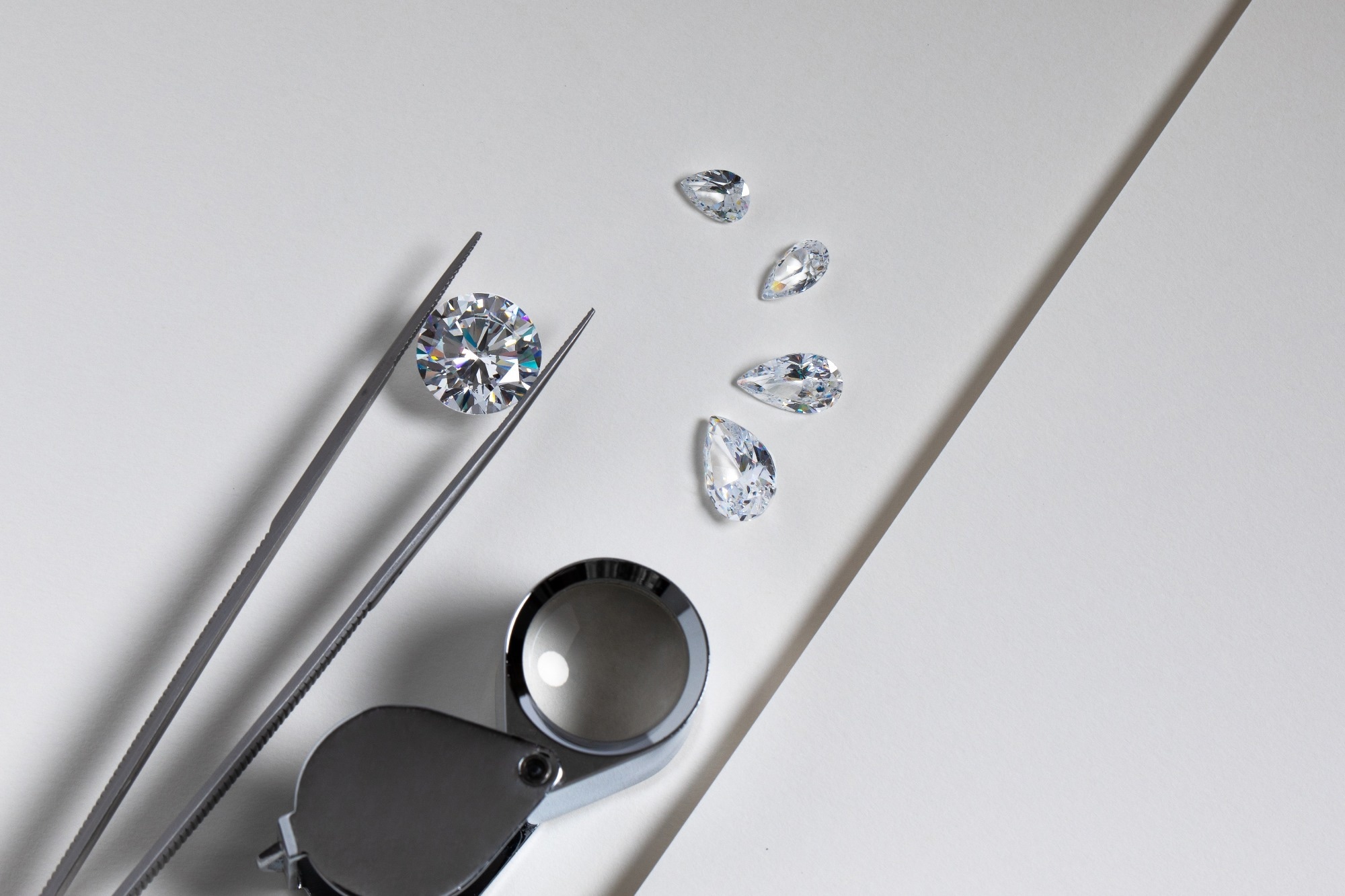
Paper: Polarization imaging for gemstone identification and metrology. Image Credit: EgolenaHK / Shutterstock
Distinguishing natural diamonds from laboratory-grown counterparts remains a significant challenge, as they share essentially the same chemical composition and crystal structure, whereas simulants such as moissanite and cubic zirconia can closely resemble diamonds despite differing compositions. A recent study accepted for publication in the journal Scientific Reports introduced a non-destructive characterization technique based on full-Stokes polarization imaging to analyze brilliant round-cut gemstones.
This method measures the degree and angle of linear polarization, as well as the degree of circular polarization, and spatial variations in these parameters provide information about crystal structure, internal stress, and growth. Researchers identified distinct polarimetric signatures in cubic zirconia, synthetic moissanite, natural diamond, and chemical vapor deposition (CVD) diamond, demonstrating the potential of full-Stokes polarization imaging for gemstone authentication and quality assessment.
Understanding Light Interaction in Gemstones
Polarization metrology examines how the polarization state of light changes as it interacts with transparent materials. When unpolarized light enters a faceted gemstone, its polarization depends on the material's refractive index, crystal structure, and internal light path. Optically isotropic materials, such as natural diamond and cubic zirconia, exhibit polarization patterns due to their facet geometry. In contrast, anisotropic materials like moissanite exhibit direction-dependent refractive indices, leading to stronger polarization effects.
These changes can be quantified using the four Stokes parameters, which describe the intensity of light and its linear and circular polarization. Full-Stokes polarization imaging generates two-dimensional maps of polarization across the entire gemstone. This allows the evaluation of crystal symmetry, internal strain, and birefringence without altering the sample.
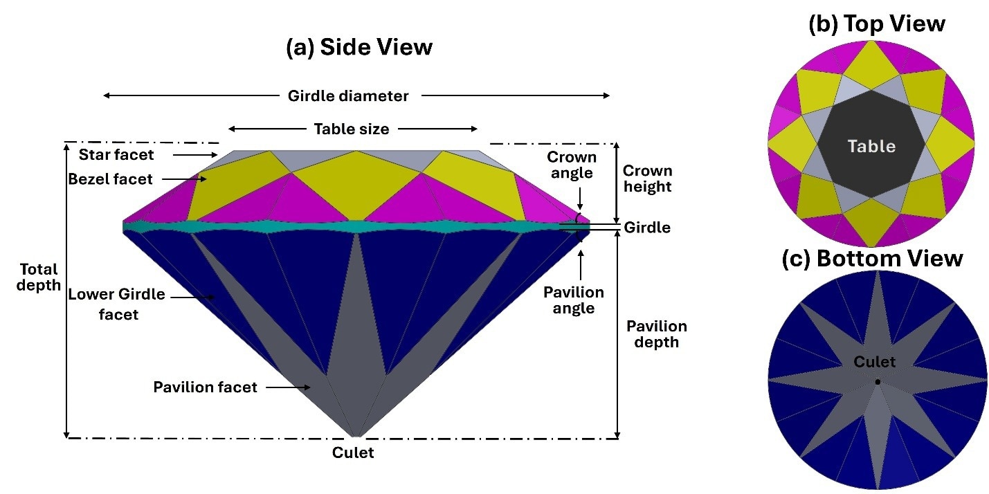
Views of a brilliant round cut show the structural anatomy of a faceted gemstone from three perspectives. (a) Side view highlights key dimensions (e.g., girdle diameter, crown height, pavilion depth) and facets (e.g., star, bezel, pavilion). (b) Top view displays the table facet and arrangement of crown facets. (c) Bottom view focuses on the culet and pavilion facets.
Methodology: Experimental Design and Equipment
To investigate the polarization response of different gemstones, researchers combined theoretical polarization ray tracing with full-Stokes imaging experiments. The optical system utilized a division-of-focal-plane polarimeter equipped with a high-resolution monochrome sensor operating across the visible spectrum. An achromatic quarter-wave plate facilitated circular polarization measurements. In contrast, a 100 mm imaging lens provided high-resolution imaging. Illumination was supplied by a broadband fiber-optic halogen source fitted with an ultraviolet-infrared (UV-IR) cut filter that restricted illumination to the visible range.
The study analyzed four round-cut gemstone materials, including natural diamond, CVD diamond, synthetic moissanite, and cubic zirconia. Two samples of each material were included. Each sample was examined in transmission, surface reflection, and side-view configurations to capture various internal light paths. The experiments generated two-dimensional maps of the Stokes polarization parameters and corresponding frequency histograms, compared with polarization ray-tracing simulations of an isotropic diamond to establish a reference for light propagation.
Distinguishing Features of Gemstone Polarization
The experiments showed clear differences in the polarization behavior of the four gemstone materials. In transmission mode, cubic zirconia exhibited a symmetric four-quadrant polarization pattern, consistent with its optically isotropic cubic crystal structure. In contrast, synthetic moissanite generated strong linear polarization due to its double refraction, producing broad high-polarization regions that distinguished it from the other materials.
The most important result was the comparison between natural and CVD diamonds. Although both share the same chemical composition and crystal structure, the samples tested showed different polarimetric signatures. In transmission imaging, natural diamonds exhibited symmetric polarization maps, strong inter-facet contrast, a well-defined secondary linear polarization peak around 0.46, and a narrow circular polarization distribution. These characteristics were consistent with a more relaxed and ordered lattice associated with natural growth.
Conversely, CVD diamonds displayed fragmented, "salt-and-pepper" polarization patterns, degraded or absent secondary linear polarization structure, with one sample showing a noisy hump near 0.40, and broader, asymmetric circular polarization distributions. These optical signatures were consistent with stress-induced anomalous birefringence associated with lattice strain during CVD crystal growth, providing a promising polarimetric signature for distinguishing natural and CVD diamond samples.
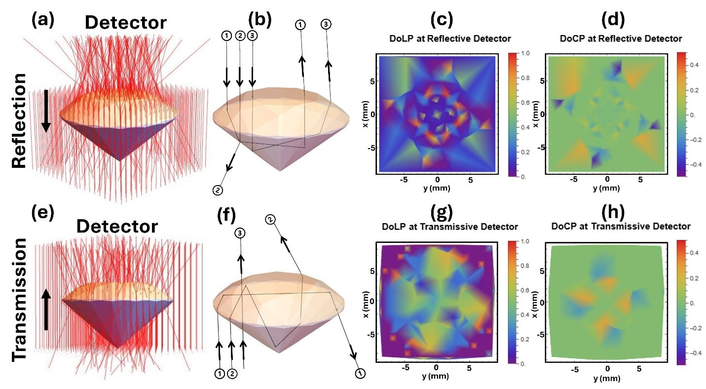
Results of polarization ray tracing for a diamond round cut with incident rays aligned along the z-axis. (a) Diagram showing the reflection of collimated rays entering the crown of the diamond. (b) Cross-sectional view illustrating the interaction of three specific rays with diamond facets. (c) The DoLP of reflected rays is shown as a spatial distribution at the reflective detector. (d) The DoCP of reflected rays is shown as a spatial distribution at the reflective detector. (e) Diagram showing the transmission (refraction) of collimated rays through the diamond. (f) Cross-sectional view illustrating the interaction of three specific rays with the diamond facets during transmission. (g) The DoLP of transmitted rays is shown as a spatial distribution at the transmissive detector. (h) The DoCP of transmitted rays is shown as a spatial distribution at the transmissive detector.
Applications in Quality Control and Material Assessment
Beyond gemstone authentication, full-Stokes polarization imaging provides a non-destructive means to assess internal optical anomalies and stress distributions in gemstones. The study presents the technique as a supplementary metrology tool for gemstone characterization and defect detection, complementing rather than replacing conventional geometric cut grading. By mapping polarization across the entire crystal, this technique can reveal stress-related spatial variations and structural irregularities that standard geometric scanners cannot directly detect.
The method may also support quality control in gemstone manufacturing workflows. Differences between cubic zirconia samples suggested sensitivity to variations in facet alignment and surface polish, providing a potential means of assessing manufacturing consistency and geometric tolerances. In gemology, the same polarimetric signatures provide an optical approach for distinguishing CVD-grown diamonds from natural diamonds, although broader validation is required before the method can be established for routine authentication.
Future Directions for Polarization Imaging Research
In summary, full-Stokes polarization imaging can convert subtle light-matter interactions into quantitative optical signatures that reveal crystal growth history, internal lattice strain, and structural quality. The observed differences between natural and laboratory-grown diamonds highlight their potential as a non-destructive tool for gemstone authentication and beyond. However, the authors cautioned that the current sample size was limited and that larger studies are needed to establish how broadly these signatures apply.
Future work should focus on refining the polarization ray-tracing framework by incorporating strain-optic tensor calculations to better represent real crystal defects. Expanding the technique to additional materials, including Type Ia and Type IIa natural diamonds, high-pressure high-temperature (HPHT) synthetic diamonds, and other crystal simulants such as yttrium aluminum garnet (YAG), gadolinium gallium garnet (GGG), and synthetic spinel, could help establish the statistical boundaries of these signatures and broaden their applications in material characterization and industrial quality control.
Disclaimer: The views expressed here are those of the author expressed in their private capacity and do not necessarily represent the views of AZoM.com Limited T/A AZoNetwork the owner and operator of this website. This disclaimer forms part of the Terms and conditions of use of this website.