The very first application of Raman spectroscopy to assess complicated biochemical changes in tumors treated with immunotherapy was achieved by researchers from the University of Arkansas.
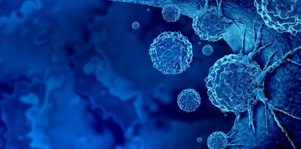
Image Credit: Lightspring/Shutterstock.com
Immunotherapy, unlike other cancer treatments, does not always result in an immediate and predicted reduction in tumor growth, and there are presently no reliable ways for determining patient response to therapy, as stated in the Scientific Reports Journal. Only a tiny percentage of patients respond to immunotherapy, and many immunotherapy combinations have serious adverse effects.
Raman Spectroscopy has been shown to be a label-free and non-destructive optical spectroscopy capable of improving diagnostic accuracy in cancer detection throughout the previous decade. Because Raman spectroscopy measurements can give a deep molecular knowledge of the biochemical alterations in cancer tissues compared to non-cancer tissues, this is the case.
Raman spectroscopic imaging was used to diagnose and grade chondrogenic tumors, including enchondroma and chondrosarcomas of escalating histologic grades, in the pilot investigation.
Raman Spectroscopy Techniques
Raman spectroscopy was used to evaluate the molecular makeup of colon cancer tumors in mice treated using two kinds of immunotherapy medicines that are now utilized in clinical treatment.
Hundreds of Raman datasets were gathered from colon cancer tumors treated with various immunotherapy treatments, and the scientists utilized machine-learning algorithms to train them.
They then compared the data from each tumor to the entire dataset to see if there was a difference between tumors that had received different forms of immunotherapy and tumors that had not.
The method successfully detected early changes in the biomolecular makeup of tumors and distinguished tumor response to different therapies. Furthermore, the researchers discovered that the alterations detected by the non-invasive Raman sensor were compatible with those revealed by comprehensive tissue analysis.
Cancer Diagnosis using Raman Spectroscopy
Cancer diagnosis continues to be one of medicine's most difficult tasks. Raman Spectroscopy (RS) is becoming increasingly important for detecting the chemical compositions of tissues and cells as new noninvasive procedures are developed or old ones are improved.
RS is a label-free and non-destructive optical technique to study basic vibrational modes of biomolecules. It has shown pathological abnormalities in the components of the bone matrices and is used to analyze the biochemical characteristics of bones. Modifications in phosphate, carbonate, and collagen breakdown, as well as spectral modifications in bone metastasis driven by prostate and breast cancer, are among these changes.
The production of calcifications was confirmed by topographical analysis, which is consistent with the histologic morphology of cartilaginous tumors, which typically reveal amorphous calcium deposits and ossification. In rare cases, excessive bone development might lead to an osteosarcoma misdiagnosis.
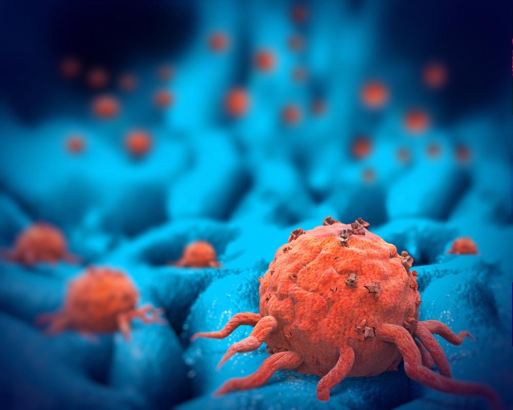
Image Credit: Giovanni Cancemi/Shutterstock.com
As a result, it is critical to determine this type of ossification and bone production by recognizing the molecular components involved. Because of the differences in clinical behavior and therapy of these malignancies, this differentiation has significant implications. The bone character of the concretions is quickly recognized by the Raman band of the phosphate group of Hydroxyapatite when using RIM (HA).
It demonstrates that optical spectroscopy may identify early changes in the biomolecular makeup of tumors with high sensitivity. This is significant because these alterations predict how well immune checkpoint inhibitors work in immunotherapy.
As a result, our research represents the first step in establishing if Raman spectroscopy may detect treatment respondents and non-responders early in the treatment process.
RS Detects Proline, Crucial Protein to Generate Collagen
RS has been proven to be a crucial instrument for studying tumor growth processes and grading in the study of the entire range of biological activity of central cartilaginous tumors of bone.
The advancement of malignancy grade appears to be highly connected to numerous biochemical contents of the ECM, such as collagen breakdown, cell proliferation, or variable biochemical composition of non-collagenous proteins in regard to proteoglycans contents.
Proline, one of the three amino acids that make up the collagen helix, is one of the biomarkers that indicates the changes and degradation of collagen in malignant tissues, according to Raman spectroscopy.
In cellular organelles, proline metabolism is critical for a variety of regulatory goals, and it is especially crucial in cancer. Because Proline serves distinct regulatory functions, a thorough understanding of its role in cancer metabolism is essential.
Apoptosis, autophagy, and the responses to food and oxygen deprivation all rely on these functions. Furthermore, Pro-derived reaction oxygen species are recognized to be a driving stimulus for cellular reprogramming. ECM is primarily generated by collagen in cartilaginous tissues, and collagen contains vast quantities of Proline that can be digested to act as a reservoir for Proline.
Cells can store or not deposit their metabolic substrates in the ECM depending on whether they are proliferating or not. Proline, on the other hand, is transformed into amino acids as collagen degrades. The RS has emphasized this process, with bands ascribed to Proline and collagen becoming less intense with time.
Malignant cartilaginous tumors are graded using a combination of histology and RS findings. The main benefits of the research method are the possibility of better diagnostic effectiveness, reduced misclassification, a precision medicine additional tool, and a significant decrease in health expenses.
References
Acunto, M., et al. (2021). Contribution of Raman Spectroscopy to Diagnosis and Grading of Chondrogenic Tumors. Scientific Reports Journal. Published: 07 February 2020. https://www.nature.com/articles/s41598-020-58848-0
Disclaimer: The views expressed here are those of the author expressed in their private capacity and do not necessarily represent the views of AZoM.com Limited T/A AZoNetwork the owner and operator of this website. This disclaimer forms part of the Terms and conditions of use of this website.