Dr. Daniele Pelliccia from RMIT University has built Australia’s most versatile X-ray instrument.
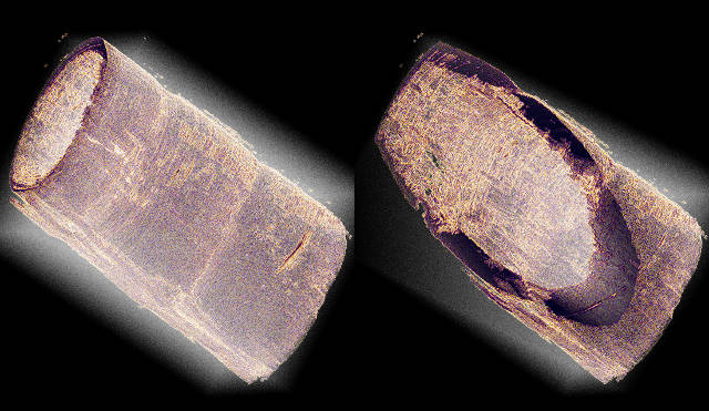 Selected 3D renderings of the micro-CT of the antenna of a tiger prawn. The specimen is about 1 mm in diameter and produces a significant attenuation for X-rays. Micro-CT enables non-destructive inspection of sample volumes. In this example both outer and inner walls as well as he structure of the antenna wall itself can be studied through virtual cuts across the rendered volume.
In conjunction with Patrik Vagovic and other researchers from Germany and the Slovak Academy of Sciences , Daniele has recently published a paper entitled “Laboratory-Based Multi-Modal X-ray Microscopy and Micro-CT with Bragg Magnifier” in the journal Optics Express (DOI: 10.1364/OE.23.018391). Using novel Bragg magnifier optics, the researchers have been able to combine different microscopy modalities together into a single instrument using a lab-sized X-ray source as opposed to resorting to synchrotron radiation.
At the heart of the system is a Rigaku FRE+ rotating anode X-ray generator high brightness X-ray source. Out of the box, the FRE+ has a focal spot size in the order of 80 to 100µm and is not generally suited used imaging applications. However, Daniele and his colleagues designed a novel optics system that magnifies the X-ray beam acting as X-ray zoom lens, resulting in an increase in resolution. While the optic is somewhat complex, it actually adds an additional level of flexibility to the system.
The optic itself consists of two channel-cut crystals in asymmetric diffraction. This produces an X-ray beam that is magnified by 15X and highly collimated that is suited to high-resolution imaging. Furthermore, thanks to the Rigaku FRE+, the system is suited to imaging weak, radiation damage sensitive samples. The Bragg magnifier optic is able to generate quantitative data for analyses such as:
- Microstructure determination
- 3D Computed Tomography or micro-CT
- Small angle scattering directionality
It performs these functions being sensitive to X-ray attenuation, refraction as well as X-ray scattering. Due to the fact that the Bragg Magnifier requires an intense X-ray beam to perform, a sealed tube type X-ray source is simply not powerful enough, nor is it able to generate high enough sensitivity.
The system is also able to operate as a multi-modal X-ray microscope. In this guise, it can image in both bright field and dark field modes, which would not be possible using a sealed tube X-ray source. This imaging mode is somewhat similar to an optical microscope and is capable of revealing high levels of detail thanks to the large variations in contrast that it can produce.
In micro-CT mode, the system is able to image samples with a 5µm resolution, which rivals other commercially available systems.
In summary, the novel optic developed by these researchers has been able to extend the imaging capabilities of the high intensity X-ray source like the Rigaku FRE+ with large focal spot to create a high resolution X-ray microscope and micro-CT. The high flux rotating anode X-ray source is key to the system, with sealed tube X-ray sources not able to produce sufficient X-ray flux to efficiently use the innovative optics to image specimens. This new experimental setup effectively brings synchrotron micro-imaging capabilities within the reach of laboratory researchers.
AXT represent Rigaku in Australia and New Zealand, distributing their XRD, WDXRF, NDT and thermal analysis products. For more details please visit www.axt.com.au or contact [email protected].