Raman spectroscopy is a powerful non-destructive analytical technique that has been widely applied in fields such as the life sciences, environmental science, and surface science. The technique is named after C.V. Raman, who discovered a previously unknown light scattering phenomenon due to the fluctuations of atoms and molecules from the normal state.
In Raman scattering, as opposed to the more common elastic Rayleigh scattering, small fractions of molecules or atoms lose or gain energy, which causes a shift in photon frequency. The energy of a molecule’s different rotational or vibrational states determines the differences in energy observed in Raman scattering. Measuring the Raman shift spectra reveals information on the structural and chemical composition of a target molecule. Essentially, this is a molecular fingerprint.
Raman spectroscopy is limited by low-intensity Raman scattering, despite advances in laser technology. Recently, the discovery of surface-enhanced Raman spectroscopy has offered significant advantages for researchers. Raman intensity can be enhanced by adsorbing molecules onto a roughened silver electrode’s surface composed of substantial amounts of nanostructured silver materials.
Research has indicated that surface-enhanced Raman spectroscopy can display improved Raman intensity by ~106. Enhancement factors as low as 107 can help realize the observation of single-molecule signals. Electromagnetic mechanism hotspots are caused by the nanogap between adjacent silver nanoparticles, significantly improving the enhancement factor of the electromagnetic mechanism, which can reach orders of magnitude higher than the chemical mechanism.
Manufacturing Stable Surface-enhanced Raman Spectroscopy Platforms
Studies have recently been performed on enhancing the stability of platforms. Research has emphasized the importance of quantitative optimization of active sites. Advances in nanomaterials, fabrication methods, and nanoscience have proven essential for this field, as better-defined nanoscale substrates can be developed.
Researchers have developed tunable zero-dimensional nanoparticles, one-dimensional nanowires, and three-dimensional arrays. Materials developed include coinage metals, MXenes, and graphene. The ability to tune the surface characteristics of nano-scaled materials has provided scientists with powerful strategies to reduce the complexities of trace detection. Active sites, each with optimal enhancement factors, can be fabricated using powerful surface characteristic modification capabilities.
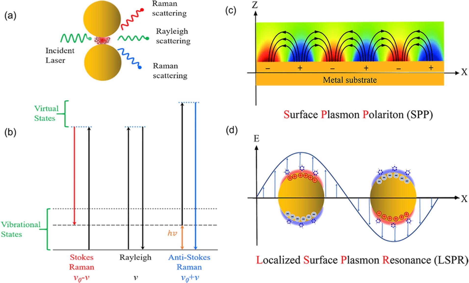
(a) Scheme of Raman and Rayleigh scattering of light by a molecule located between two metallic nanoparticles involving a hot spot. (b) Jablonski diagram representing the quantum energy transitions for Raman and Rayleigh scattering of a molecule. Schematic diagrams illustrating (c) surface plasmon polaritons at the surface of metal thin film and (d) localized surface plasmon resonance at metal nanoparticles. Image Credit: Long, L et al., ACS Materials AU
The Study
The authors have presented a review of the dimensional design of substrates used for enhancement purposes in surface-enhanced Raman spectroscopy. Whilst the authors have not provided a truly comprehensive review of studies from the past half a century, they have attempted to outline the development of these substrates.
The research has investigated zero-dimensional, 1D, 2D, and 3D substrates categorized according to their geometric dimension. For each of these substrate classes, composite configuration and geometric design can be modified to achieve the optimal enhancement factor at their active sites.
Finding an ideal substrate for this analytical technique is important. Substrates must possess several beneficial properties such as high sensitivity, selectivity, and stability, which will make them attractive for both research and technological applications. Possessing these optimal characteristics improves the versatility of the technique, improving the enhancement factor, fast response, and enhanced signal reproducibility.
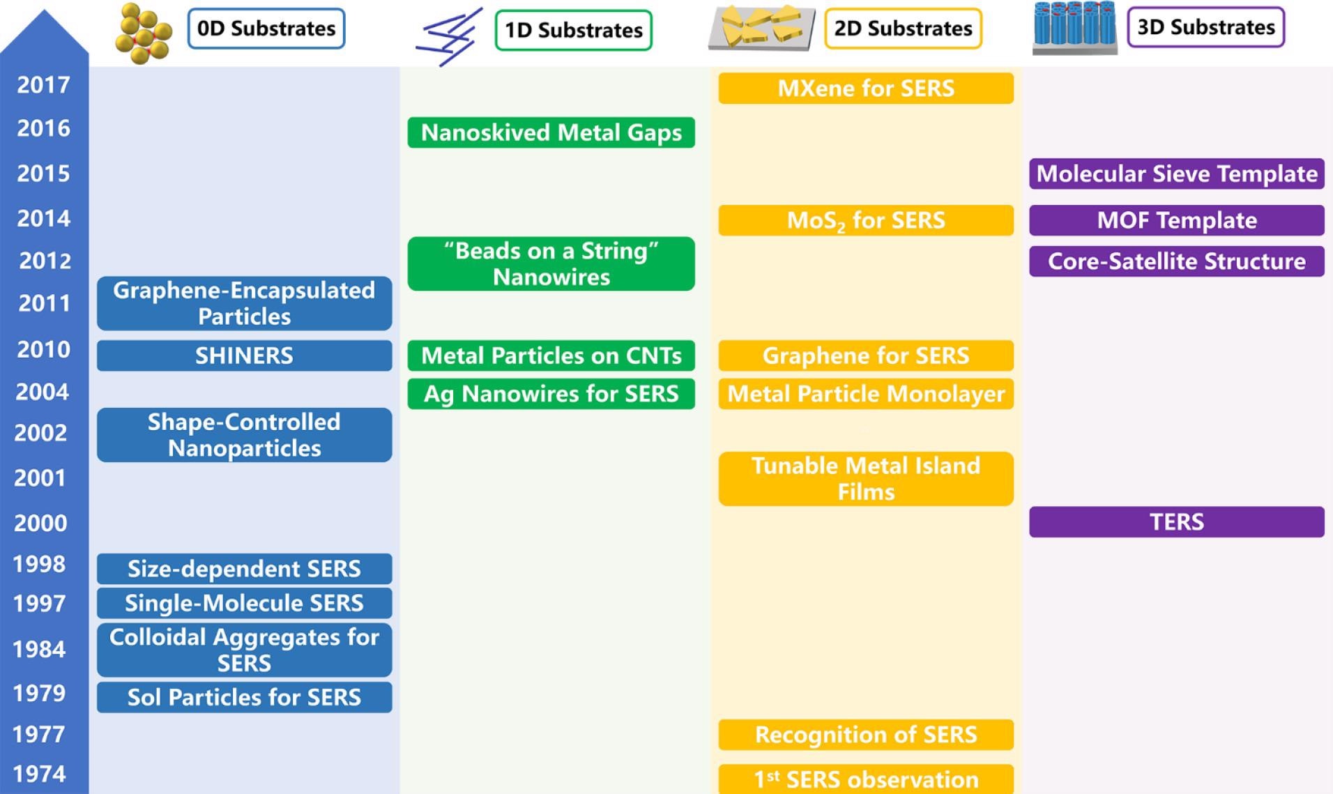
Some milestones in the development of SERS substrates made in the past nearly half-century history of SERS, including 0D nanoparticles, 1D nanowires, 2D metallic and nonmetallic thin films, and 3D nanostructure arrays. The advances of SERS substrates have significantly broadened the application scope of SERS. Image Credit: Long, L et al., ACS Materials AU
4D Surface-enhanced Raman Spectroscopy
In the review, the authors have highlighted that femtosecond pulse laser technology can be employed to incorporate a temporal dimension into surface-enhanced Raman spectroscopic analysis. By incorporating a temporal dimension into this analytical technique, surface-enhanced Raman spectroscopy can provide information on the ultrafast dynamics that occur when molecules undergo structural change. This capability broadens the scope of the technology beyond providing information on the chemical composition of molecules.
This technique is referred to as 4D surface-enhanced Raman spectroscopy and provides unprecedented information on target molecules. The authors have stated that this added spatiotemporal resolution helps the technique answer fundamental questions and enhances its versatility as an ultrafast analytical technique.
Providing ultrafast analysis of nanometer-resolution structures and single molecules is the ultimate goal of surface-enhanced Raman spectroscopy research. Currently, however, there are no suitable techniques that can achieve this. Observing molecular change dynamics and visualizing specific important chemical reaction dynamics is currently beyond the reach of researchers.
The dimensional design may help to advance spatiotemporal resolution for this analytical technique and enable quantitative single-molecule analysis. A 4D design where the pump-probe process is carefully planned, in accordance with enhancing a substrate’s geometric structures, may be able to meet this demand. Furthermore, incorporating strategies such as scanning probe methods may help to realize this goal of surface-enhanced Raman spectroscopy.
The Future
Whilst there are still significant challenges to address in the field, this review paper provides a wealth of information on current progress and advances in these techniques, and the authors have stated that in the future it will be possible to visualize elementary chemical reactions and achieve four-dimensional analysis of molecules and their ultrafast dynamics.
Further Reading
Long, L et al. (2022) Dimensional Design for Surface-Enhanced Raman Spectroscopy ACS Materials Au [online] pubs.acs.org. Available at: https://pubs.acs.org/doi/10.1021/acsmaterialsau.2c00005
Disclaimer: The views expressed here are those of the author expressed in their private capacity and do not necessarily represent the views of AZoM.com Limited T/A AZoNetwork the owner and operator of this website. This disclaimer forms part of the Terms and conditions of use of this website.