Cryo-electron microscopy (cryo-EM) has advanced rapidly in recent years, enabling atomic-level structural analysis of viruses and proteins through Single Particle Analysis (SPA) workflows. These applications demand highly stable hardware and software systems. To support this progress, JEOL introduces its latest cryo-TEM: the CRYO ARM™ 300.
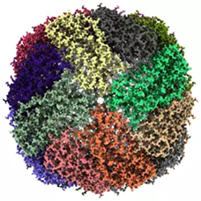
Image Credit: JEOL USA, Inc.
The CRYO ARM™ 300 delivers exceptional resolution and stability, enabling fully automated, unattended image acquisition for SPA. As shown in the figure, Fujita et al. achieved a 1.29 Å structure of mouse apo-ferritin using the CRYO ARM 300 at SPring-8 (Japan).
Automated Specimen Exchange System
The system features an autoloading specimen stage cooled to liquid nitrogen temperatures, along with a cryo-storage device capable of long-term storage for up to 12 frozen-hydrated specimens. Liquid nitrogen is automatically supplied to both the stage and the storage unit as needed.
The autoloader supports loading and retrieving up to four grids at once, without disturbing the remaining samples in storage. A unique labeling and tracking system ensures specimen management is seamless and transparent to the operator. These capabilities make beamline-style scheduling of microscopy practical and efficient.
Cold Field Emission Gun (Cold FEG)
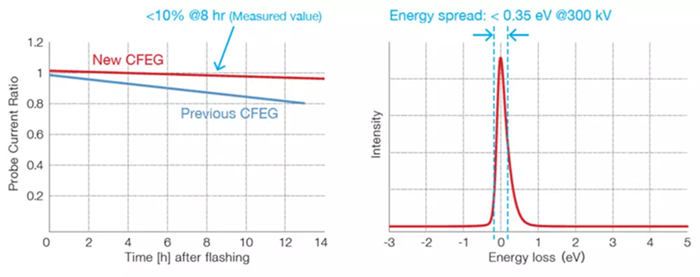
Image Credit: JEOL USA, Inc.
The cold field emission gun (FEG) generates a high-brightness electron beam with an exceptionally small energy spread, providing excellent temporal coherence. It also enhances spatial coherence, allowing the CRYO ARM™ 300 to deliver superior resolution and contrast compared to any other 300 kV cryo-TEM currently available.
In-Column Energy Filter (Omega Filter)
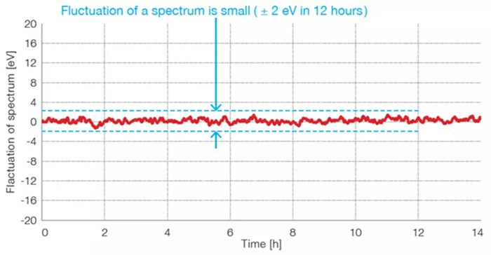
Image Credit: JEOL USA, Inc.
Equipped with an enhanced in-column energy filter (Omega filter), the CRYO ARM™ 300 can acquire both energy-filtered images and energy loss spectra. Zero-loss imaging provides higher contrast by minimizing chromatic aberration.
The filter’s stability has also been significantly improved (see figure), allowing Single Particle Analysis runs to require only two adjustments of the zero-loss peak position over a 24-hour period. A new Auto Adjust function simplifies the setup process, ensuring that users of all experience levels can easily configure the filter for optimal data acquisition.
Single Particle Analysis Image Acquisition Software
The CRYO ARM™ 300 is compatible with both the JEOL Automated Data Acquisition System (JADAS) and SerialEM (UC Boulder).
Hole-Free Phase Plate*1
The hole-free phase plate (HFPP) consists of a thin, continuous carbon sheet placed in the back focal plane of the objective lens. This results in a significant improvement in image contrast of frozen-hydrated specimens. The HFPP is manufactured using a unique technology, which results in strong and reliable performance at cheap prices.
Zero Fringe Illumination*2
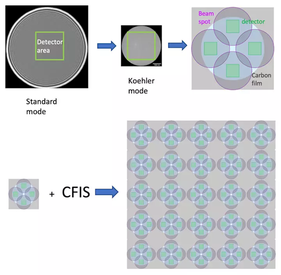
Image Credit: JEOL USA, Inc.
The CRYO ARM™ 300 can be equipped with Köhler illumination, which significantly reduces beam-edge fringes. This allows for the use of a narrow, parallel electron beam when capturing images using Coma-Free Image Shift (CFIS) or beam/image shift, whether within a single hole during SPA runs or even across multiple holes on a quantifoil grid (see figure).
Auto Adjustment Functions*3
The system includes a suite of automated adjustments - such as auto focus, auto coma-free alignment, and auto parallel-beam illumination - ensuring image acquisition is consistently performed under optimal conditions.
*1 & 2: Optional unit. *3: Images acquired with the bottom mount camera are used.
Key Features
High Throughput - Increased Productivity for Cost-Effectiveness
Quick Collection
The JEM-3300 CRYO ARM™ 300 II offers high-speed data acquisition by integrating beam shift and precision specimen stage movement. This system's superior beam control reduces beam tilt, which causes coma aberration, even with beam shift. As a result, image quality can be maintained while throughput is enhanced.
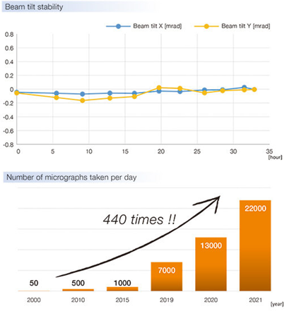
Image Credit: JEOL USA, Inc.
Zero Fringe System
The CRYO ARM™ 300 II offers a distinct “Koehler mode” illumination, in addition to the standard beam. This mode has no interference fringes, eliminates electron-beam damage to non-imaging areas, and allows users to capture more images in a smaller area.
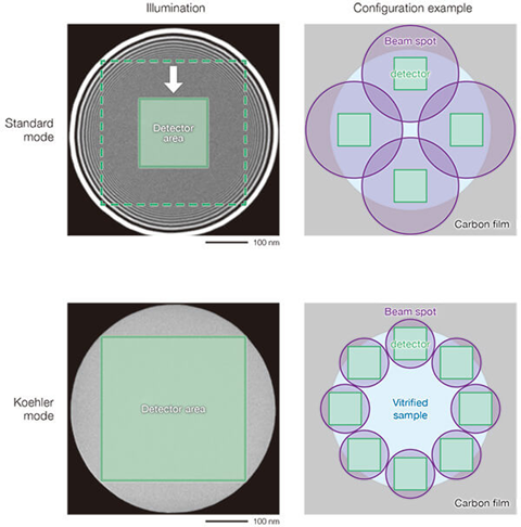
Image Credit: JEOL USA, Inc.
Highly Flexible Operation
High Precision Stage Drive
The CRYO ARM™ 300 II features a rotation-free stage, offering excellent positional reproducibility. This allows users to move samples between the microscope column and cryo-storage without losing alignment, making it possible to reuse low-magnification images, referred to as the "global map." As a result, optimal sample conditions can be maintained while significantly minimizing electron beam exposure, helping to ensure the acquisition of high-quality data.
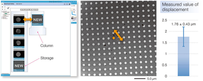
Image Credit: JEOL USA, Inc.
JADAS 4
The CRYO ARM™ 300 II comes with a new version of "JADAS<sup>*</sup>" software, allowing beginner users to capture data. The new JADAS allows users to automatically capture image data by simply specifying the acquisition parameters through a wizard.
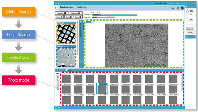
Image Credit: JEOL USA, Inc.
Contamination Free Sample Storage And Removal
Grids can remain clean in storage for weeks or longer inside the CRYO ARM™ 300 II. Additionally, individual grids can be replaced using a transfer magazine, allowing for efficient handling without disturbing the remaining samples. Throughout specimen ejection, liquid nitrogen temperatures and a nitrogen gas atmosphere are maintained to preserve sample integrity.
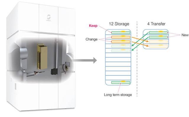
Image Credit: JEOL USA, Inc.
High Stability
New Cold Field Emission Electron Gun
The CRYO ARM™ 300 II comes with a new cold field emission electron gun (CFEG). This new gun not only has a low energy spread, but it also has increased probe current stability, allowing users to capture high-quality images at all times.
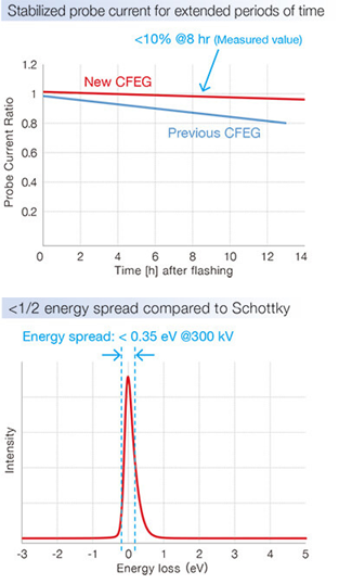
Image Credit: JEOL USA, Inc.
New Omega Filter
The CRYO ARM™ 300 II is equipped with a newly improved in-column energy filter, the Omega filter, which features a self-adjusting mechanism to consistently deliver optimal performance with high stability.
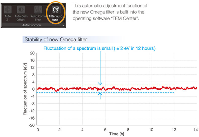
Image Credit: JEOL USA, Inc.
Specifications
Main instrument. Source: JEOL USA, Inc.
| |
|
| Electron gun |
Cold field emission gun (New CFEG) |
| Standard accelerating voltage |
300 kV, 200 kV |
| Energy filter |
In-column Omega energy filter (New Omega Filter) |
| Accessory |
Hole-free phase plate |
Specimen stage / Automated specimen exchange system / Specimen storage. Source: JEOL USA, Inc.
| |
|
| Specimen stage |
| Coolant |
Liquid nitrogen, Automated liquid-nitrogen filling system built-in |
| Specimen cooling temperature |
100 K or less |
| Temperature measurement |
Near specimen, Cryo-shield, Near LN2 tank |
| Specimen movements |
| X, Y |
Motor drive (Movements: ± 1 mm)
Piezoelectric elements |
| Z |
Motor drive (Movements: ± 0.2 mm) |
| Tilt-X |
Motor drive ( Tilt: ± 70°) |
| Rotation |
Settable to 0°or 90° within the specimen plane |
| Specimen exchange system |
Air-lock, Automated cryo-transfer system built-in |
| Cooling temperature |
105 K or less |
| Specimen exchange cartridge |
Up to 4 specimens can be changed at one time. |
| Specimen storage |
Up to 12 specimens can be held |
| Coolant |
Liquid nitrogen, Automated liquid-nitrogen filling system built-in |
| Cooling temperature |
105 K or less |
Options
- Automated tilt series acquisition software for MicroED
- Bright-field STEM detector
- Dark-field STEM detector
- Direct detection camera
- EDM (Electrostatic Dose Modulator)
- JADAS (JEOL Automated Data Acquisition System for Cryo-EM)
- Low accelerating voltage
- TEMography
Gallery
A GroEL structure at 1.98 Å resolution was achieved using just 504 micrographs - a significant improvement compared to the 3.1 Å resolution obtained from 1883 micrographs in a previous study (as of October 26, 2020, EMDB).
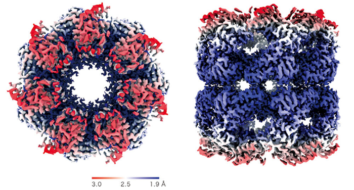
Image Credit: Data courtesy of Dr. Junso Fujita at Osaka University..
A cryo-electron micrograph (left), 3D density map (center), and fitted atomic model (right) of human hemoglobin were obtained using high-speed data collection at a rate of 850 movies per hour.
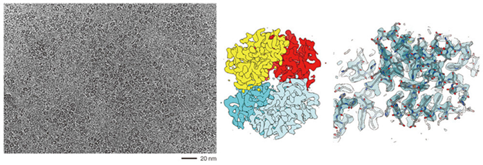
Image Credit: Specimen courtesy of Dr. Miki Kinoshita at Osaka University.
2.5 Å resolution maps of the photosystem I trimer from Acaryochloris marina are shown below, viewed from the stromal side perpendicular to the membrane plane (left) and from the side along the membrane plane (right).
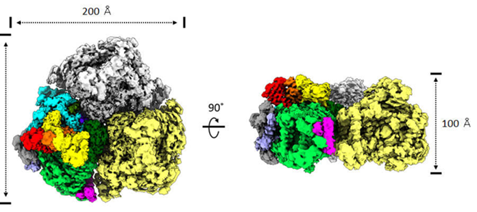
Image Credit: Data courtesy of Dr. Koji Yonekura at RIKEN, Spring-8 Center.
An electron diffraction pattern of a thin catalase crystal, acquired using the Omega filter, shows clear diffraction spots extending to approximately 2.1 Å along the diagonal axis.
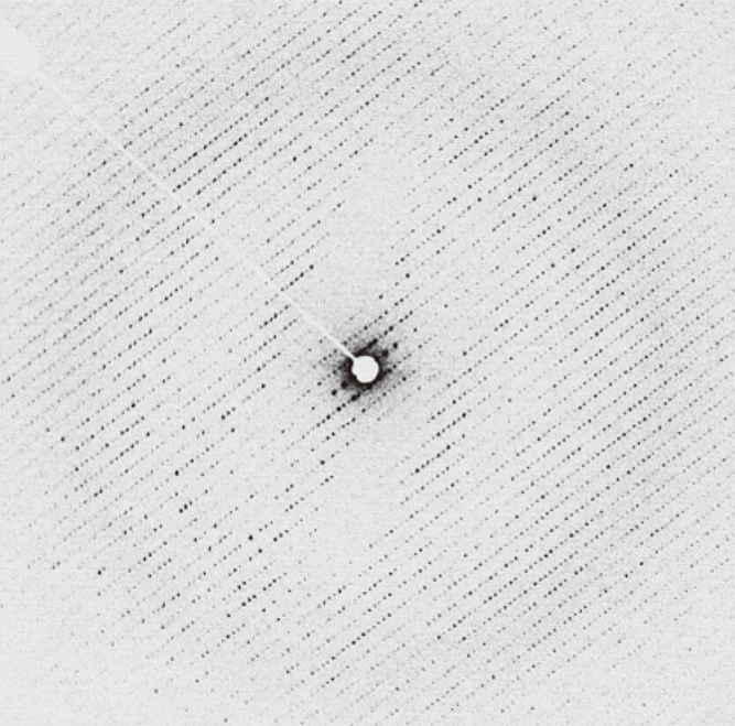
Image Credit: Data courtesy of Dr. Koji Yonekura at RIKEN, Spring-8 Center.
A cryo-electron tomogram of isolated exosomes was acquired using a hole-free phase plate. The tomogram was reconstructed from micrographs collected over a tilt range of –60° to +60°, in 2° increments.
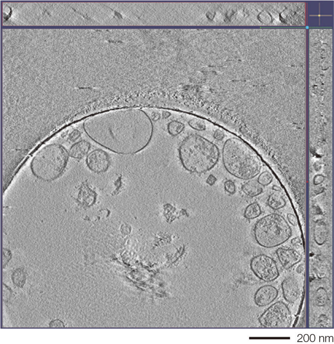
Image Credit: Specimen courtesy of Dr. Naoomi Tominaga at National Cancer Center Research Institute and The University of Tokyo.