Dark contamination areas can gradually develop on the specimen’s surface during electron microscope observation. This contamination may obscure the true surface structure or degrade resolution in affected regions.
The 'Dark Square' phenomenon occurs during electron beam microscopy when the electron beam irradiates hydrocarbon-based impurities on the specimen's surface, causing carbonaceous deposits. The Excimer UV Cleaner uses UV light (Xe excitation at 172 nm) to remove these contaminants from the specimen surface. This method can also be used to hydrophilize a specimen’s surface.
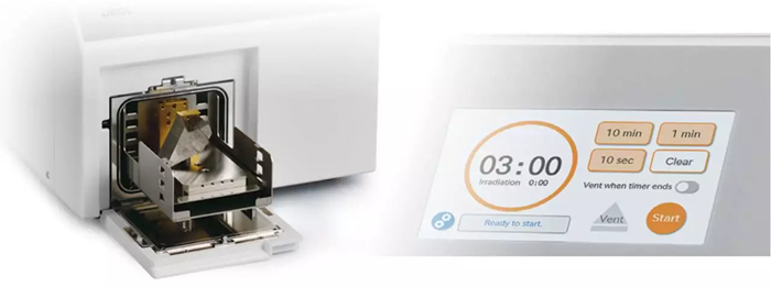
Image Credit: JEOL USA, Inc.
Key Features
- Minimizes contamination during electron microscope observations.
- Cleans and purifies specimens and tools.
- Improves the hydrophilicity of the specimen surface.
Mitigate Contamination During Observation
Imaging at low accelerating voltage is essential in SEM to reveal delicate surface structures in greater detail. However, surface contamination can obscure the observation of this fine structure.
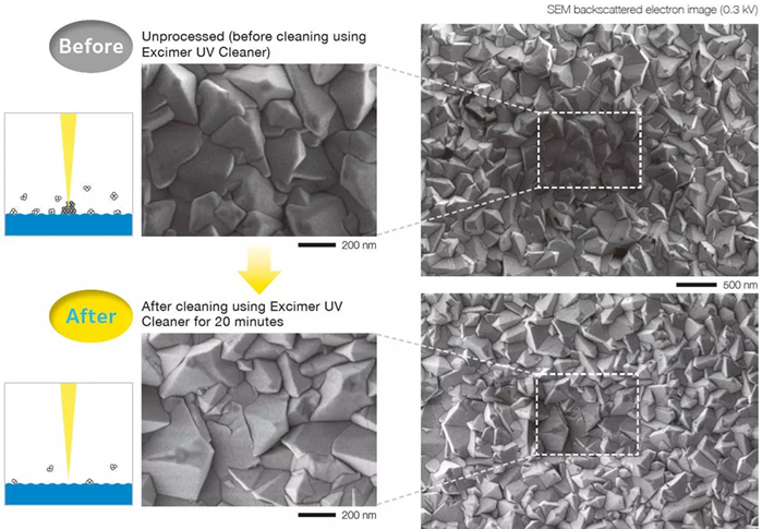
Image Credit: JEOL USA, Inc.
The observations were conducted at a magnification of 100,000 (left image) and 30,000 (right image). In the untreated material (top row), the area imaged at 100,000 times appears darker and less defined.
However, after cleaning with the Excimer UV Cleaner (bottom row), contamination induced by electron beam irradiation was reduced, as seen by the reduced number of hydrocarbon compounds on the surface.
Cleaning Contamination Adhered to the Specimen Surface
Contamination accumulates over time when a specimen is handled or maintained in an environment containing organic chemicals.
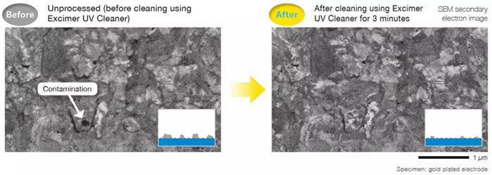
Image Credit: JEOL USA, Inc.
The Excimer UV Cleaner can remove organic substances from a dry atmosphere.
Hydrophilization
Reactive oxygen species are produced when the Excimer UV Cleaner is exposed to ultraviolet light. These can form polar functional groups on the surface, thereby increasing hydrophilicity. This pre-treatment procedure can increase bonding when preparing a material for EM observation.
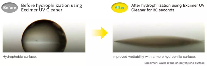
Image Credit: JEOL USA, Inc.
Mechanism for Specimen Cleaning / Hydrophilization Using the Excimer UV Cleaner
Process of Contamination Caused by the Electron Beam
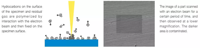
Image Credit: JEOL USA, Inc.
Cleaning Process
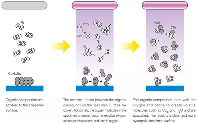
Image Credit: JEOL USA, Inc.
The specimen tray height can be adjusted to accommodate larger specimens.
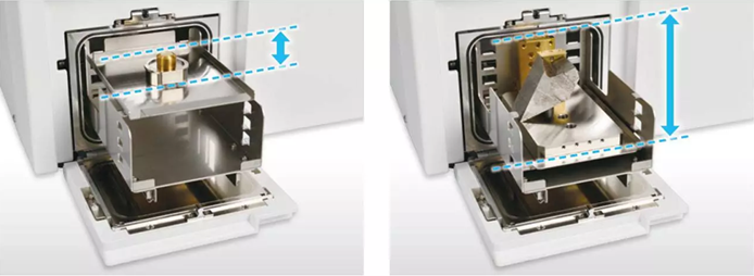
Image Credit: JEOL USA, Inc.
Specifications
Source: JEOL USA, Inc.
| |
|
|
|
| Wavelength of irradiated UV light |
172 nm |
| Maximum specimen size |
105 mm (W) × 105 mm (D) × 70 mm (H) |
| Maximum specimen mass |
1 kg |
| Irradiation time |
10 seconds to 99 minutes |
| Vacuum pump |
Diaphragm-type dry vacuum pump |
| Installation requirements: |
Power |
|
Single phase 90 to 110 V AC, 50 / 60 Hz, 500 VA |
Dimensions and weight
(cables and vacuum pipes not included) |
Main unit |
39 cm (W) × 50 cm (D) × 32 cm (H), approximately 22 kg |
| Vacuum pump |
50 cm (W) × 20 cm (D) × 30 cm (H), approximately 9 kg |
| Room temperature |
15 to 30 ℃ |
| Humidity |
60 % (RH) or less (no condensation) |
| Regular replacement parts (replace them every time the cumulative irradiation time reaches 1000 hours) |
UV lamp, vacuum gauge, O-ring, etc. |