The JSM-IT710HR Field Emission SEM is a small, multipurpose Schottky Field Emission SEM that offers the newest intelligent technology for imaging and analysis with high spatial resolution.
This microscope is perfect for high-resolution applications because of its innovative in-lens field emission gun and sophisticated electron optics, which allow it to deliver large probe currents while keeping the probe small.
Despite its small size, this extremely adaptable SEM has a large chamber and High and Low Vacuum modes for handling a wide range of specimen types in their natural state.
Smart – Flexible - Powerful
Smart
The most recent advancements in JEOL's intelligent technology make it accessible at any level. Clear, high-resolution images are produced in a matter of seconds by best-in-class auto functions, which include alignment and focus.
Zeromag's workflow is quick, using the integrated optical camera for navigation and a smooth transition to SEM imaging. The analytical models can be utilized to view live EDS X-ray and spectrum maps.
They have advanced it further by adding automation from Montage (large-area mosaics) to Simple SEM for automatic image collection at various locations, magnifications, and conditions. All of this technology is crammed into a small platform for unmatched usability.
Flexible
The JSM-IT710HR has a sizable specimen chamber with several ports ideally positioned for analytical attachments such as heating/cooling sub-stages, multiple EDS, EBSD (co-planar with EDS), WDS, CL, STEM, etc.
The large, internal, mechanically eccentric stage makes it simple to position and align heavy, large specimens before shutting the door and leaving the chamber.
Powerful
High spatial resolution imaging and analytical results are produced by combining JEOL's special in-lens field emission gun, which can emit beam currents of up to 300 nA, with an aperture angle control lens that maximizes large probe currents to the smallest probe diameter.
Thanks to the high-sensitivity, quadrant BSE detector's live 3D surface reconstruction feature, users can view specimens with complex topography more clearly. JEOL's fully integrated EDS system for Real-Time, Live EDS spectra, and Live X-ray maps are examples of analytical models.
Built-in automation streamlines and improves throughput, and internal data management software connects all data for a real-time view of analysis locations. This SEM is open to Python scripting and allows remote control and live web viewing.
Key Features
- The in-lens specimen receives ≥300 nA from the Schottky field emission gun.
- Sophisticated auto features such as focus, astigmatism correction, and beam alignment
- Numerous ports in a large specimen chamber
- The chamber is equipped with a large, mechanically eucentric specimen stage.
- Zeromag makes navigation easier by enabling a smooth transition between optical and SEM images.
- Every piece of data is connected: SEM images, color images, and EDS data for a map of every analysis site.
- Live Analysis: Complete JEOL EDS integration with live spectrum and live X-ray map automation in real time.
- Automation. Montage: Automate EDS maps (analytical models) and multiple large area image mosaics (stitching). Simplify workflow and automate routine imaging with Simple SEM. Recipes for detectors, magnifications, multiple kV, and more.
- Models for both high and low vacuum for any kind of sample. Simple one-click switching between high and low vacuum.
- Specimen Exchange: A comprehensive, integrated guide that covers everything from the introduction of the specimen to the automatic creation of images. Unmatched usability at any level.
- Minimal environmental impact and low maintenance needs (no cooling water needed).
Zeromag
Simple navigation between the optical and live SEM images.
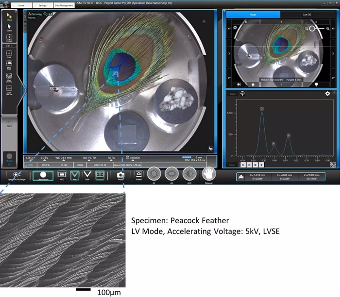
Image Credit: JEOL USA, Inc.
Real-Time EDS – Live Analysis
JEOL EDS Embedded for Live Analysis. View the Live Map or Live Spectrum. With a single click, switch between views.
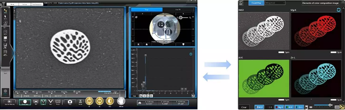
Image Credit: JEOL USA, Inc.
Live 3D
For samples with complicated topography, the Quadrant Backscatter Detector can show a live 3D image, which adds more information.
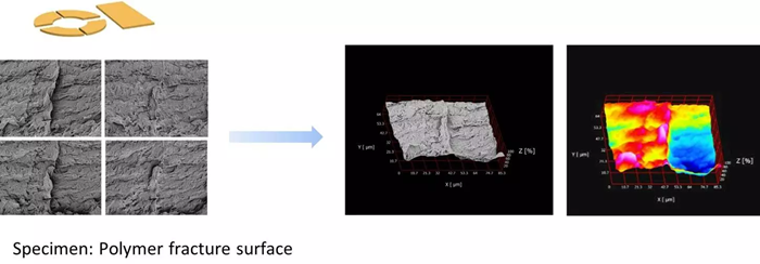
Image Credit: JEOL USA, Inc.
Automation – Montage (Automatic Large Area Image Capture)
Automatically take pictures and stitch them together to create a montage. Contains analytical models and X-ray maps.
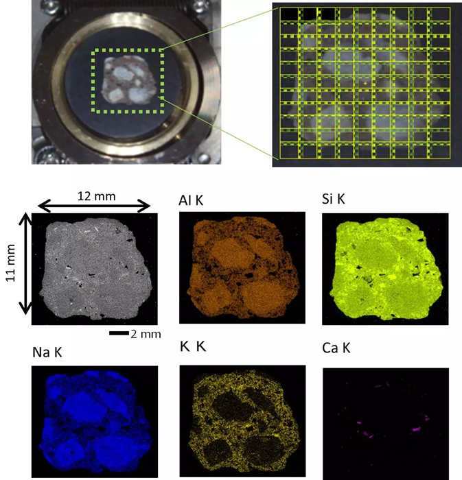
Specimen: Limestone, number of fields:99 (9 × 11), Accelerating voltage: 12 kV, Magnification/field: X70. Image Credit: JEOL USA, Inc.
Automation – Simple SEM for Image Capture
Automate the gathering of images under various circumstances, at various locations, and various magnifications:
- Set Conditions
- Set Target Areas
- Start
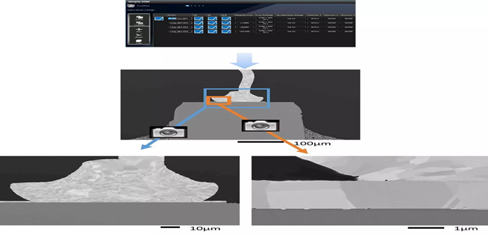
Image Credit: JEOL USA, Inc.