The high-sensitivity, low-noise 19 MP CMOS sensor delivers clearer imaging, allowing fine specimen details to be observed, even under low electron dose conditions.
With a global shutter and high frame rate (58 fps in full-pixel mode), it enables image series acquisition with fewer artifacts during dynamic observations.
The "SightX" camera control software offers intuitive, user-friendly operation for streamlined workflows.
Key Features
- Digital Zoom ensures sharp images even at lattice resolution.
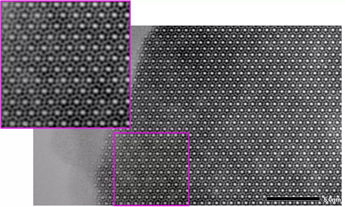
High resolution TEM image of Si3N4. Instrument: JEM-2100Plus. Accelerating voltage: 200 kV. Image Credit: JEOL USA, Inc.
- Digital Zoom allows for the observation of atomic configurations that exhibit quasi-periodicity, such as those found in quasicrystals.
- The great dynamic range produces outstanding electron diffraction pattern contrast, from the high-intensity direct spot to the lower-intensity spots.
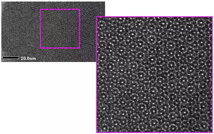
High-resolution TEM image and electron diffraction pattern of Al72Fe24Ni4 dodecagonal quasi-crystal. Instrument: JEM-2100Plus. Accelerating voltage: 200 kV. Image Credit: JEOL USA, Inc.
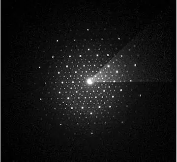
A bright triangle area appearing at upper right of the diffraction pattern is shown by Log display. Image Credit: JEOL USA, Inc.
- Fine structures, such as twin textures of polycrystalline silicon, can be seen in high contrast. See the below TEM images of polycrystalline silicon.
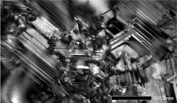
BF-TEM. Instrument: JEM-2100Plus. Accelerating voltage: 200 kV. Image Credit: JEOL USA, Inc.
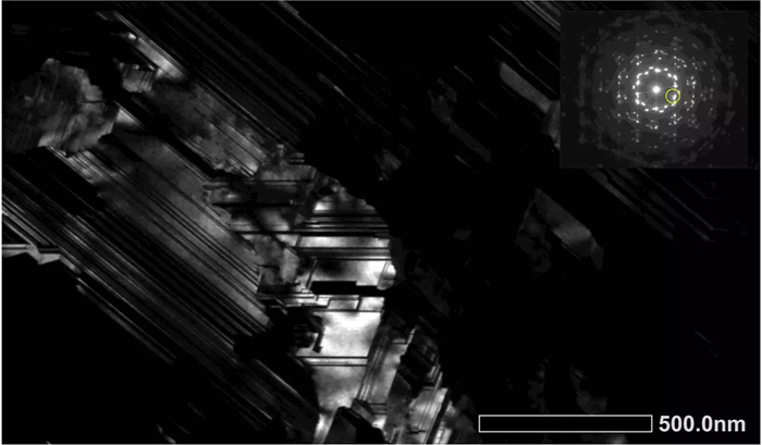
DF-TEM. Dark field image formed by the diffraction spot highlighted with the yellow circle in the inset. Image Credit: JEOL USA, Inc.
- Digital Zoom allows for clear examination of lamella formations. See the TEM images of polyethylene (PE) and polypropylene (PP) resins below.
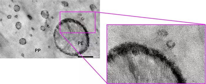
Sample preparation method: Microtome. Instrument: JEM-2100Plus. Accelerating voltage: 80 kV. Image Credit: JEOL USA, Inc.
- Membrane structures of chloroplasts and mitochondria can be detected, and tiny membrane features can be studied in depth. See the TEM image of carrot leaves below.
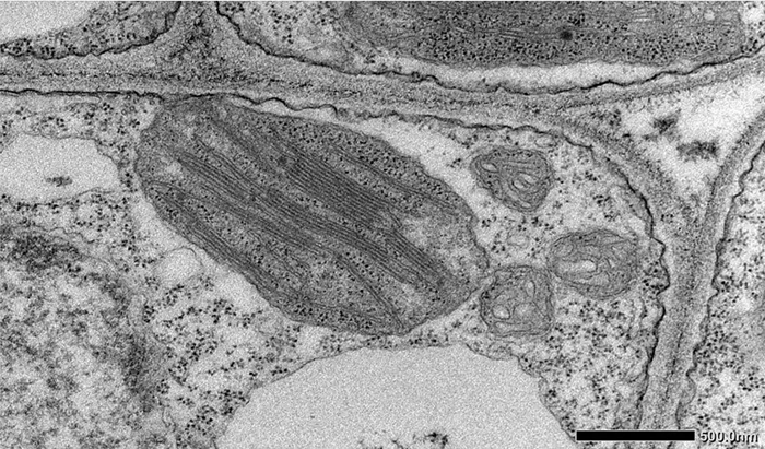
Image Credit: JEOL USA, Inc.
- Liposome lipid bilayer membranes are readily distinguishable, as seen in the TEM image of the phospholipid bilayer (liposome) below.
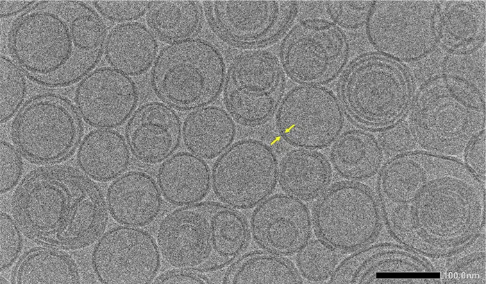
Sample preparation method: ice embedding. Instrument: JEM-F200Cryo. Accelerating voltage: 200 kV. Image Credit: JEOL USA, Inc.
- Camera system control software “SightX”.
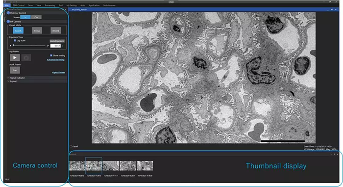
Image Credit: JEOL USA, Inc.
Specifications
Source: JEOL USA, Inc.
| . |
. |
| Accelerating voltages |
≦ 200 kV |
| Number of effective pixels |
19 M pixels (5,688 × 3,336 pixels) |
| Sensor active size |
36.40 mm × 21.35 mm |
| Pixel size |
6.4 μm × 6.4 μm |
| Frame rate |
58 fps / All pixel mode |
| Recording modes |
Image, Video (Stack frame) |
| Image formats |
L size (5,688 × 3,336), M size (2,880 × 1,680), S size (1,248 × 1,200) |
| File formats |
TIFF (Image, Video), BMP (Image), JPG (Image) |
| Shutter type |
Global shutter |
| Mounting position |
Bottom mount (below viewing chamber), non-retractable |
| Control Software |
SightX or TEM Center (JEM-F200 and CRYO ARM™200(JEM-Z200FSC)) |
Applicable models: JEM-F200, JEM-2100Plus, JEM-1400, JEM-2100, JEM-2100F, JEM-2200FS, CRYO ARM™200 (JEM-Z200FSC)
* SightSKY uses a novel high density feedthrough connector – transferring sensor signals and providing power supply between the vacuum chamber and atmospheric electrical unit - adopting the patent, No.JP6635605, of RIKEN (Institute of Physical and Chemical Research) that is manufactured by MIST and Lead Electronics Co. Ltd.