The Oxford Instruments witec360 redefines Raman imaging and correlative microscopy. It provides verstaile nanoscale analysis to fulfill the requirements of researchers in academia and industry.
The witec360 Raman microscope, the successor to the alpha300 series, introduces new standards for performance and broadband capability. Its modular design provides immediate and long-term benefits. This design supports easy adoption for new Raman spectroscopy users and offers the flexibility needed for diverse and evolving applications.
Benefits
Acquire Research-Grade Results
Optimised optics throughout the entire beam path deliver enhanced speed, sensitivity, and resolution.
Experience Unprecedented Versatility
Broadband capabilities provide spectral flexibility for demanding research tasks and diverse applications in multi-user facilities.
Gain Comprehensive Insight
The seamless combination of analytical methods enables the correlation of chemical and structural characteristics, resulting in a more complete sample analysis.
Configure the Microscope for Current and Evolving Experiments
A modular design offers extensive capabilities and upgrade options, enabling tailored solutions and scalability to match individual requirements and resources.
Enhance Productivity with Intelligent Automation
Advanced hardware automation and intuitive software streamline data acquisition and analysis, improving operational efficiency and facilitating rapid user onboarding for individuals with various experience levels.
Ensure Consistency and Transparency
Reproducibility is enhanced, and compliance with institutional and regulatory requirements is supported through multi-user management, internal calibration, and automated reporting.
Advanced Confocal Raman Technology for Every Task
- Time-resolved measurements to track reactions and material alterations
- 2D area scans for high-resolution imaging and fast overview mapping
- 3D analyses that disclose internal structures layer by layer
- Point-by-point spectral acquisition for precise measurement at individual locations
- Line scans for depth profiling along defined axes
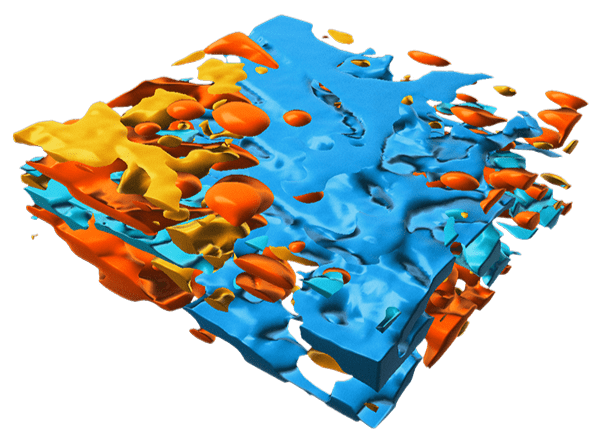
3D Raman image of a cosmetic emulsion rendered in Imaris. Dimensions: 30 x 30 x 10 µm3. Image Credit: Oxford Instruments
Modularity
Flexible – Sustainable – Future-Proof
The witec360 system is built upon a modular architecture. This allows for the precise customisation of configurations to address diverse experimental challenges.
The system's offerings encompass a spectrum of solutions that range from fixed setups optimised for single applications to specialised instrumentation for advanced academic research.
The design's inherent flexibility ensures the witec360's capacity for evolution and expansion along with experiments.
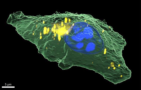
3D Raman image of a human epithelial cancer cell rendered in Imaris. Sample courtesy of Dr. Irina Estrela-Lopis and Tom Venus, Institute of Medical Physics and Biophysics, Leipzig University, Germany. Image Credit: Oxford Instruments
Explore the witec360
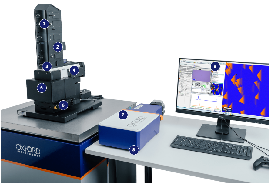
Image Credit: Oxford Instruments
1. Modular Input and Output Couplers
- The system supports individual combinations of imaging modes and correlative techniques.
- It allows for optimal alignment to ensure an ideal beam path and high throughput.
- Includes polarisation-maintaining optics.
- Offers manual and automated variants.
- Features the RayShield coupler and RayLine filters for superior low-frequency signal detection.
- A polarisation module is available for freely rotating the polarisation direction or generating circularly polarised light.
2. Lasers
- Utilises fiber-coupled lasers to ensure maximum flexibility and efficiency.
- Offers extensive options for wavelength and power range.
- Allows for up to six wavelengths to be permanently coupled within a single system.
- Incorporates high-precision fiber couplers for rapid switching between different lasers.
- Features patented TruePower™ technology for absolute laser intensity control.
- Includes automatic shutter control.
- Equipped with polarisation-maintaining optics.
3. Illumination and Detection Setup
Flexible illumination and detection features accommodate different research fields and sample shapes.
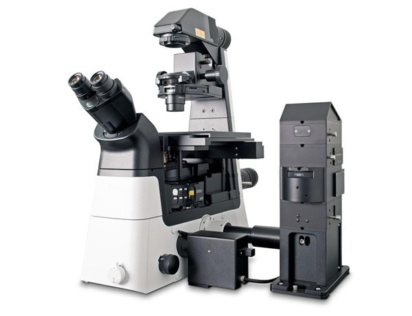
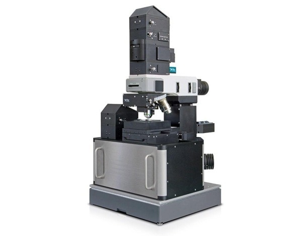
- The microscope body can be upright or inverted for illumination from above or below.
- BeamFlex offers optimal flexibility for upright and inverted imaging, including reflection and transmission configurations.
4. Whitelight/Widefield Microscopy
- Utilises class-leading Köhler illumination to ensure uniform, high-contrast images across the entire field of view.
- Delivers high-end white light microscopy images across multiple modes: Brightfield, Darkfield, and DIC (Differential Interference Contrast).
- Capable of operating in either reflection or transmission mode.
- An optional polarisation setup is available.
- Optional integration for fluorescence microscopy provides multi-modal imaging capability.
5. Objective Turret
- The system allows for up to six objectives to be mounted simultaneously.
- It is compatible with high-performance objectives from various suppliers.
- Features an optional software-controlled turret rotation for increased workflow efficiency.
6. Sample Positioning and Handling
Sample Positioning
- Ideal for precise scanning in X, Y, and Z axes. Both manual and motorised stages are available.
- Piezo stages for applications demanding the highest spatial resolution and precision.
- Available with standard travel ranges from 25 mm × 25 mm up to 350 mm × 300 mm
Sample Handling Accessories
- Includes stages designed to accommodate special experimental conditions.
- Supports environmental control devices such as heating and cooling stages and stage-top incubators.
- Provides sample holders compatible with various devices and sample geometries.
7. Spectrometers
Hexalight
- Mirror-free, lens-based design maximises signal detection.
- The device, accommodating up to six gratings, delivers peak resolution and adaptable use, removing the necessity for manual configuration.
- Proprietary Olaf-Hollricher-Design (OHD) lenses offer true broadband performance from 350 to 1100 nm.
- Integrated device allows seamless switching between high-resolution and broadband acquisition of Raman and PL spectra.
- Up to 6 installed gratings provide both high resolution and application flexibility, with automated selection via an innovative harmonic drive.
- Available in 300 mm or 600 mm focal lengths.
Monolight
- The system utilizes a single, fixed grating solution, making it optimised for specialised applications.
- Offers configuration options to accommodate lasers with wavelengths ranging from Ultraviolet (UV) at 266 nm to Near-Infrared (NIR) at 1064 nm.
Detectors
- Utilises ultra-sensitive Oxford Instruments Andor cameras. These cameras are optimised for advanced Raman imaging.
- Ultra-low dark current
- Excellent quantum efficiencies
- Ultra-fast acquisition times
- Improved imaging quality by reduced artifacts
- Offers a range of options from budget-conscious to high-end detectors (e.g., Front Illuminated, Back Illuminated, Electron Multiplying, Open Electrode).
- Includes InGaAs array detectors to provide extended possibilities within the Near-Infrared (NIR) excitation range.
8. Compact Footprint
- The system allows for a flexible layout of all components.
- It ensures reduced heat and vibrational interference during measurements.
- The design maintains compatibility for operation within a glove box or enclosure.
9. Software
- Features an Integrated Software Suite that manages both data acquisition and evaluation.
- Built with a modular architecture to allow for adaptable software configurations.
- Includes an optimized user-interface for powerful instrument control.
- Provides seamless control of supported correlative techniques.
- Offers intelligent data analysis and visualisation capabilities
Advanced Software Packages
- ParticleScout: Enables fast and easy Raman particle identification and classification.
- TrueMatch™: Includes Raman spectral database management software for simplified compound identification.
- Workflow Manager: Facilitates automated and routine measurement workflows.
- Multi-user Management: Allows for precise control of data access and instrument functionalities for individual users and user groups
- Programming Interface: Supports custom-defined measurement procedures for individualised applications.
- DaVinci: Serves as an accurate nanomanipulation and nanolithography tool
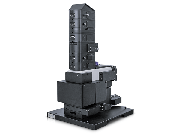
Upright. Image Credit: Oxford Instruments
Inverted. Image Credit: Oxford Instruments
BeamFlex. Image Credit: Oxford Instruments
Correlative Microscopy
Comprehensive Sample Analysis with Raman and Correlative Techniques
The witec360 platform can incorporate correlative methods to yields comprehensive insights into chemical, topographic, mechanical, and structural characteristics.
The platform's associated software offers streamlined operational management and facilitates automated data correlation.
Upgrade options for correlative imaging techniques are available. These upgrades can be applied to current witec360 systems. They are also available for legacy alpha300 series microscopes.
Correlative Imaging Techniques
Scanning Electron Microscopy (SEM)
SEM techniques provide essential nano-structural, crystallographic, and elemental insights for materials analysis, nanotechnology, and geosciences.
Image Credit: Oxford Instruments
Photoluminescence (PL) Imaging
Photoluminescence (PL) is a widely used technique in materials science and semiconductor characterisation. It offers critical insights into optoelectronic properties.
Image Credit: Oxford Instruments
Atomic Force Microscopy (AFM)
AFM provides crucial information on nanoscale surface properties, including topography, adhesion, and stiffness. These insights are valuable for materials science and nanotechnology applications.
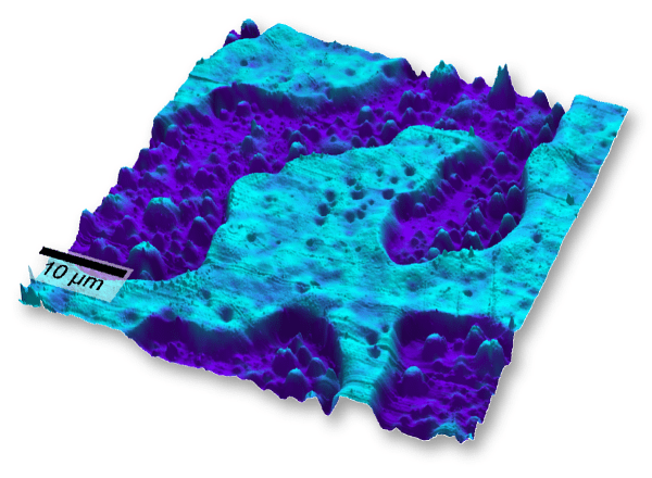
Image Credit: Oxford Instruments
Topographic Raman Imaging
TrueSurface enables simultaneous topographic and Raman measurements. This is particularly useful for samples with irregular surfaces, such as rocks, tablets, or micro-structured materials.
Image Credit: Oxford Instruments
White-Light Microscopy Options
Köhler illumination in witec360 microscopes ensures optimal white-light images. This facilitates efficient location and visualisation of regions of interest.
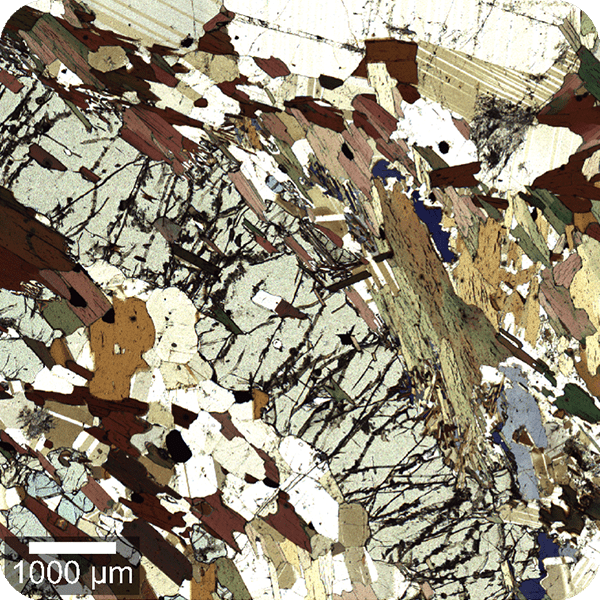
Image Credit: Oxford Instruments
Scanning Near-Field Microscopy (SNOM)
Scanning Near-field Optical Microscopy (SNOM) achieves optical imaging with resolutions as fine as approximately 60 nm.
Image Credit: Oxford Instruments
Fluorescence Microscopy
For life science applications that utilise fluorescent labeling, Raman data can be complemented by integrating fluorescence microscopy.
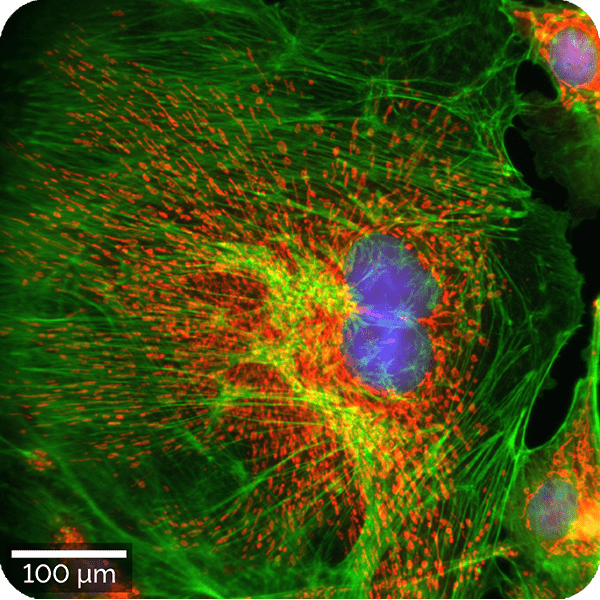
Image Credit: Oxford Instruments
Time-Correlated Single Photon Counting
Time-Correlated Single Photon Counting (TCSPC) techniques, such as TLM and FLIM, are especially useful in semiconductor, optoelectronics, and life sciences research.
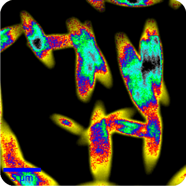
Image Credit: Oxford Instruments
Antibunching Measurements
For quantum computing and cryptography, single-photon emitters (SPEs) are identified using correlative Raman, Photoluminescence (PL), and antibunching techniques.
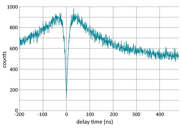
Image Credit: Oxford Instruments
Second Harmonic Generation (SHG)
SHG is very sensitive to non-centrosymmetric structures. It is effective for analysing the properties of crystals within 2D materials and enabling label-free investigations in the life sciences.
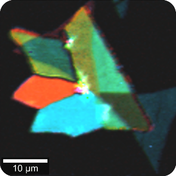
Image Credit: Oxford Instruments
Performance
Class-Leading Performance
The witec360's components are optimised for superior transmission efficiency and consistency. This ensures rapid and precise data acquisition. Their design streamlines imaging workflows and enables the successful execution of complex experiments.
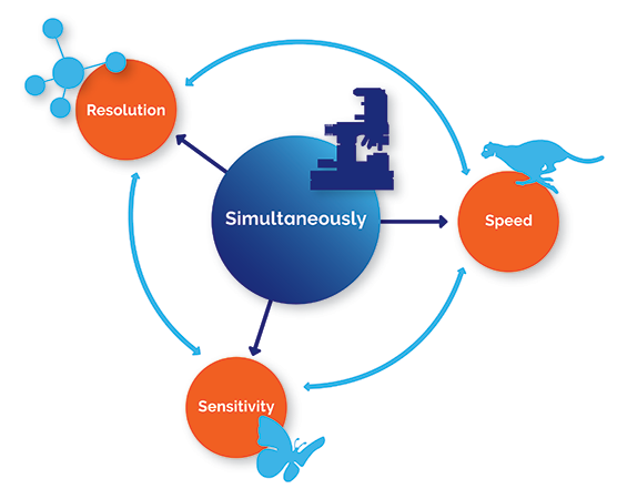
Image Credit: Oxford Instruments
Maximised Signal Intensity for the Broadband Range
- 350 - 1100 nm within the same core system
- Extended options from UV (266 nm) to NIR (1064 nm)
Diffraction-Limited Spatial Resolution
- Lateral resolution < 300 nm (dependent on excitation wavelength)
- Depth resolution < 950 nm (dependent on excitation wavelength)
Exceptional Spectral Resolution
Resolution is achieved down to 0.1 cm⁻¹ relative wavenumbers (at 633 nm excitation)
Ultra-Fast Raman Imaging
Spectral acquisition occurs at rates of less than 1 millisecond
Hexalight - the Core of witec360’s Performance
The witec360 utilises the advanced Hexalight spectrometer. This spectrometer features lens-based, mirror-free optics and six grating positions to maximize throughput and spectral adaptability.
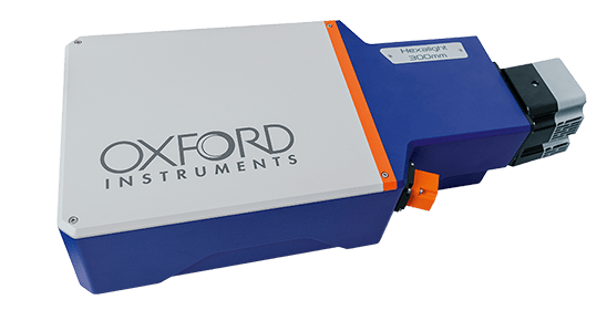
Image Credit: Oxford Instruments
Automation
Effortless Precision With Advanced Automation
witec360 microscopes offer advanced automation features to boost efficiency and streamline processes. These features contribute to consistent and reliable data acquisition.
Key Benefits:
- Ease of use: Simplifies intricate processes and allows for advanced measurements, accommodating both beginner and experienced Raman imaging users.
- Streamlined workflow: Automated adjustment processes boost efficiency, allowing more time for research activities.
- Enhanced reproducibility: Guarantees dependable, high-quality outcomes via automated management of routine tasks and hands-free optics alignment.
- Remote operation: Enables full remote control of measurements and microscope operation, ideal for environmnetal chambers such as glove boxes.
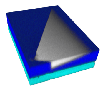
3D Raman image of a defect in a SiC wafer. Dimensions: 180 x 245 x 7 µm3. Image Credit: Oxford Instruments
Automation for Every User
Multi-User Labs:
This approach supports diverse skill sets, simplifying processes with easy-to-use shared equipment.
Industry Researchers:
It executes repetitive experimental procedures rapidly and accurately, speeding up progress.
Raman Newcomers:
Complex tasks are made easier, allowing for reliable use with straightforward training.
Raman Experts:
Explore new possibilities using sophisticated automation for intricate experiments.
witec360 Automation Key Features
Instrument Setup and Imaging Control
| Feature |
Benefit |
|
Autobeam – One Click Precision Alignment
This system provides automatic laser beam path alignment. It uses a certified reference sample for diffraction-limited resolution and stable performance. The system includes a memory function for various laser alignments and supports full remote operation.
|
The system is designed for multi-user laboratories and enclosed setups. It offers hands-free, repeatable alignment. This ensures consistent, high-quality outcomes.
|
|
TrueCal – Calibration Made Easy
Automated, pre-configured calibration routines are included. These routines ensure the system remains spectrally aligned and prepared for experiments. This makes the system suitable for repetitive or regulated workflows.
|
This makes the system suitable for repetitive or regulated workflows. These workflows require precision, compliance, and quantitatively accurate results.
|
Raman Imaging
| Feature |
Benefit |
|
TrueComfort – Simplify Everyday Operations
Essential operations are streamlined to enhance convenience and accelerate imaging.
- Motorised Köhler illumination optimises light settings
- Quick switching between bright-field and Raman modes
- The objective revolver rotates with a single click via software control
|
A cost-effective approach created to improve productivity and streamline standard imaging tasks.
|
|
TrueSurface – Always in Focus
It ensures consistent focus throughout the measurement process, resulting in clear, high-quality images.
|
It is optimised for large-area scanning and is suitable for samples with intricate shapes, such as curved, tilted, and structured surfaces.
|
|
Motorized Laser Coupler – Expand the Wavelength Options
Software-controlled selection of up to six laser wavelengths, spanning the UV to NIR range.
|
Suitable for multi-wavelength experiments and a wide variety of samples requiring diverse excitation settings.
|
|
TruePower™ – Absolute Laser Power Control
Absolute laser power is precisely controlled throughout measurements, ensuring consistent and reproducible illumination with an accuracy better than 0.1 mW (extending to 0.1 µW in advanced configurations).
|
This functionality is indispensable for energy-dependent experiments and for sensitive samples requiring stringent laser power regulation.
|
|
Motorized Polarization Modules – Effortless and Accurate
The automated modification of freely rotating excitation polarisers and detection analysers streamlines and speeds up polarization-dependent and polarisation-resolved series analyses.
|
Improve the study of molecular orientation and anisotropic materials by enhancing usability and accelerating workflows.
|
|
Motorized and Piezo-Driven Stages – Precision Made Easy
Attain accurate, automated sample positioning with integrated motorised and piezo-driven scan stages.
|
This feature is essential for thorough large-area mapping, automated focusing on specific areas, and precise re-evaluation of previous measurement locations. |
Data Processing
Utilise the advanced software for streamlined data handling and examination, facilitating swift result comprehension.
| Feature |
Benefit |
|
Software Suite Analysis Wizard
Data analysis is simplified via guided, click-through workflows.
|
User training time decreases, and data analysis is expedited for all experience levels.
|
|
TrueComponent Analysis
Sample components within an image are identified and differentiated automatically.
|
Complex samples experience rapid and thorough analysis, with spectral unmixing simplified.
|
|
TrueMatch
Measured spectra are quickly matched to known molecules using established spectral databases.
|
Direct peak assignment occurs without interpretation or mathematical assessment.
|