Using larger microspheres, the Wide Field-of-View SMAL effectively doubles the field of view in comparison to traditional SMAL designs. This improvement comes with a decrease in resolution and magnification, which make it best suited to biological imaging applications.
Product Codes:
BIOL-001-OW-M25 → M25 thread, 60 mm parfocality
BIOL-001-OW-RMS → RMS thread, 45 mm parfocality
Key Features
- Enhanced Field of View: Roughly double that of traditional SMAL lenses
- Resolution Trade-Off: A slight decrease in resolving power and magnification as a result of increased microsphere use
- Optimal for Bio-Imaging: Especially effective for the imaging of mammalian cells and comparable biological specimens
Resolution
Spatial resolution down to 100 nm demonstrated using semiconductor samples.
Intended Applications
- Imaging of microchips or similar small-scale structures
- Life sciences, cells
Specifications
Source: LIG Nanowise Limited
| Attribute |
Description |
| Field of View (FOV) |
30 μm |
| Resolution / Magnification |
100 nm/180X |
| Ideal Environment |
Immersion (water or oil) |
| Use Case Focus |
Biological imaging, especially where larger areas need scanning |
| Thread type |
RMS or M25 x 0.75 mm |
Application Examples
- Biological Preference: More suitable for scanning cells or tissues where the field size is more significant than achieving the absolute highest resolution
- Design Trade-Offs: As the field of view (FOV) expands, both resolution and magnification diminish; this is a crucial factor for targeted applications
- Integration: Compatible with current microscope systems
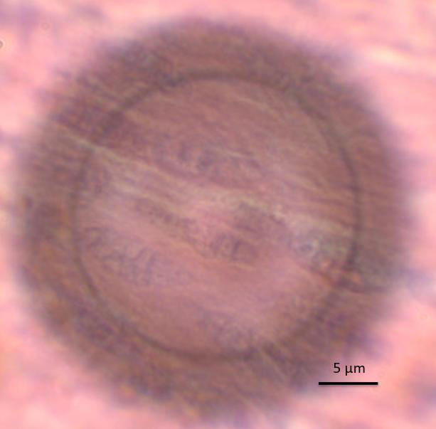
Mammal intestine cells imaged using a wide-field-of-view SMAL lens. Image Credit: LIG Nanowise Limited
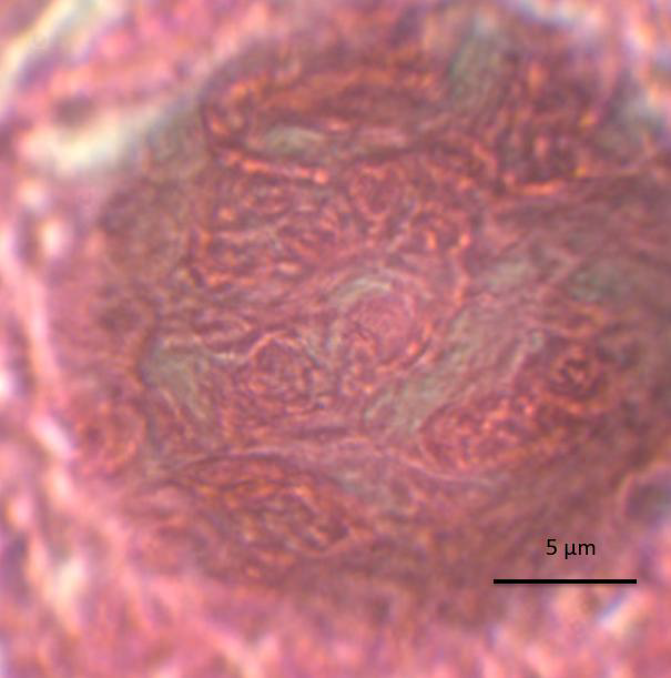
Mammal ovary cells imaged using a wide-field-of-view SMAL lens. Image Credit: LIG Nanowise Limited
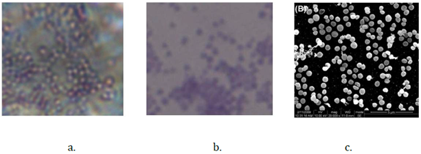
a) Staphylococcus aureus imaged with SMAL; (b) conventional 100× objective; (c) TEM. Image Credit: LIG Nanowise Limited