The HT7800 series comes in three variants: HT7800, HT7820, and HT7830 (see Specifications table below). These microscopes mark a new era of transmission electron microscopy, combining Hitachi’s 75+ years of expertise with modern digital innovations. Designed for both experienced researchers and newcomers, the series delivers high-resolution imaging with a user-friendly interface and ergonomic controls that improve efficiency and comfort.
Developed for life sciences, materials science, and industrial applications, the HT7800 series offers flexible imaging modes, high-speed digital processing, and seamless automation to ensure accurate, reproducible results. The microscopes can also be operated under normal room lighting, eliminating the need for a darkroom and enhancing usability and comfort.
- Enhanced digital imaging system: With low-dose functionality for improved sample integrity
- Expandable capabilities: STEM, EDX, MirrorCLEM, and other specialized accessories for different research needs
- Dual-Mode objective lens: Switch between high-contrast mode and high-resolution mode with a single click for unmatched flexibility
- User-friendly interface: Modern, intuitive GUI allows seamless operation in normal room light conditions
Features and Benefits
Dual-Mode Objective Lens for Maximum Versatility
- Quickly switch between high-contrast and high-resolution modes on a single microscope
- Eliminates the need for multiple instruments, optimizing workflows and lowering costs
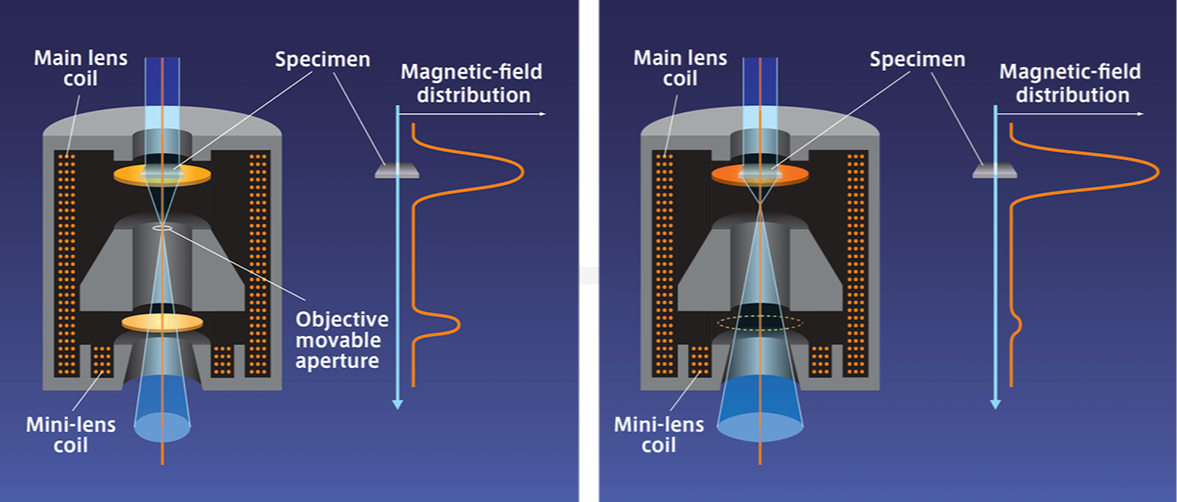
Image Credit: Hitachi High-Tech Europe
Intuitive Operation and User-Friendly Interface
- The HT7800 series offers an intuitive digital GUI suitable for both beginners and experienced users
- Operate in a well-lit environment with the integrated CMOS screen camera, reducing fatigue and improving ergonomics.
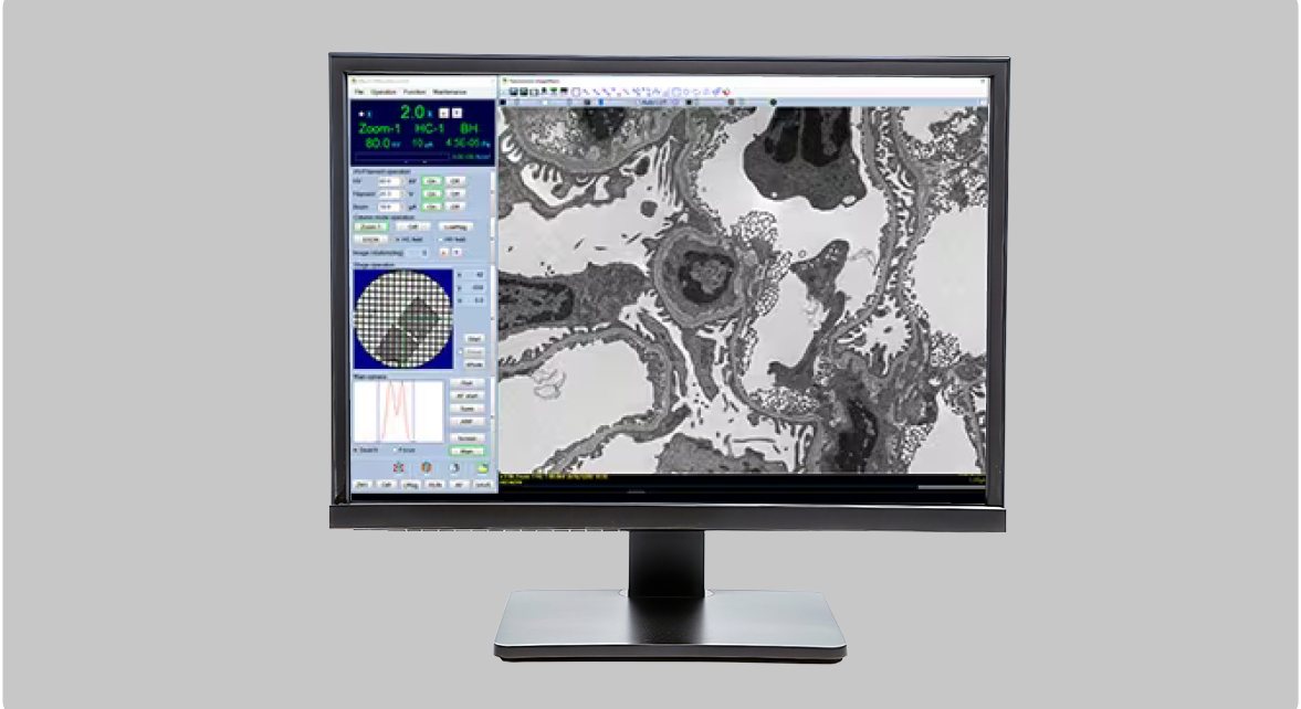
Image Credit: Hitachi High-Tech Europe
The HT7800’s Digital Imaging System
- A high-speed CMOS camera for superior imaging
- Whole View Function enables fast, automated image acquisition over large areas
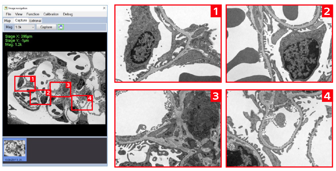
Image Credit: Hitachi High-Tech Europe
Automation and Navigation
- Auto Multiple Frame (AMF) Imaging: Seamlessly stitch multiple images together for high-resolution panoramic views
- Advanced image navigation & mapping: Quickly locate, track, and analyze regions of interest
- 3D electron tomography: Acquire tilted images to generate accurate 3D reconstructions
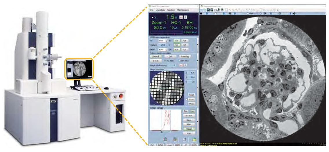
Image Credit: Hitachi High-Tech Europe
Expandable and Customizable for Advanced Research Needs
- STEM and EDX: For elemental mapping and material characterization
- MirrorCLEM: Integrates fluorescence microscopy with TEM imaging for more comprehensive research analysis
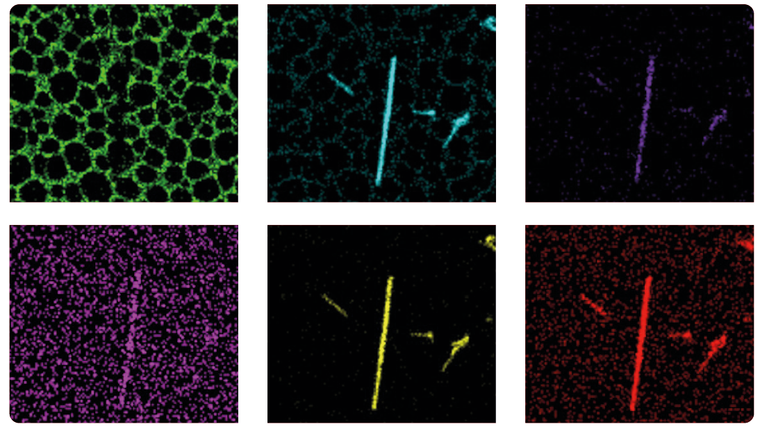
Image Credit: Hitachi High-Tech Europe
Specifications
Source: Hitachi High-Tech Europe
| |
HT7800 |
HT7820 |
HT7830 |
| Electron gun |
W (standard), LaB6 |
LaB6 (standard), W |
LaB6 (standard), W |
| Accelerating voltage |
20 - 120 kV (100 V/step variable) |
20 - 120 kV (100 V/step variable) |
20 - 120 kV (100 V/step variable) |
| Resolution (Lattice) |
0.20 nm (Off-axis, 100 kV) |
0.14 nm (Off-axis, 120 kV) |
0.14 nm (Off-axis, 120 kV)
0.19 nm (On-axis, 120 kV) |
| Maximum magnification |
x600,000 |
x800,000 |
x1,000,000 |
| Stage maximum tilt angle |
±70° |
±30° |
±10° |
Standard
features |
Auto focus, Microtrace, Autodrive, Live FFT display, Measurement function, Low dose, API (auto pre-irradiation),
Image navigation function, Column with mild baking function, Whole view function, Drift correction function, etc. |
Applications Gallery
Life Sciences and Biomedical Research
- High-contrast imaging for biological specimens
- Low-dose mode for cryo-TEM and delicate biological samples
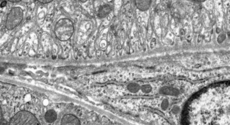
The images below show a comparison between a conventionally stained segment and an unstained section utilizing the HT7800 series High-Contrast lens.
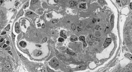
Mouse Kidney (stained), accelerating voltage 80 kV, magnification x300. Image Credit: Hitachi High-Tech Europe
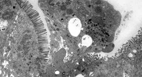
Rat jejunum (unstained), accelerating voltage 80 kV, magnification x2,000. Image Credit: Hitachi High-Tech Europe
Material Science and Nanotechnology
- High-resolution imaging of nanoparticles, polymers, and advanced materials
- STEM & EDX compatibility for compositional analysis
The image below highlights hollow-cone dark-field observation using an Au/TiO2 catalyst on the HT7820. The electron beam diffraction region used for the hollow-cone dark-field observations is indicated in yellow within the selected-area electron diffraction pattern. TiO2 diffraction waves can be clearly observed in the hollow-cone dark-field images.
Au/TiO2 catalyst. Bright-field TEM image, accelerating voltage 120 kV. Image Credit: Hitachi High-Tech Europe
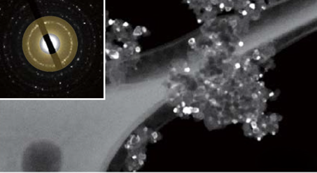
Au/TiO2 catalyst. Hollow-cone dark-field TEM image, accelerating voltage 120 kV. Image Credit: Hitachi High-Tech Europe
Particle/Polymer
- High-resolution performance and digitization allow you to quickly observe the sequence of several nm pores.
The images below show TEM observation results of mesoporous silica particles expected to be applied to drug delivery systems.
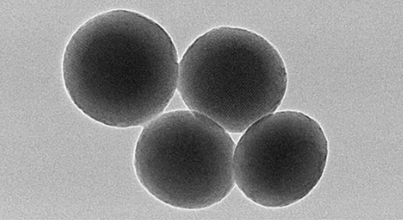
Mesoporous silica powder, accelerating voltage 120 kV, magnification x70,000. Image Credit: Hitachi High-Tech Europe
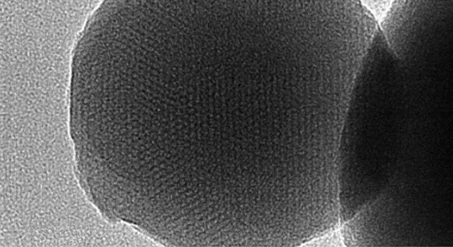
Mesoporous silica powder, accelerating voltage 120 kV, magnification x200,000. Image Credit: Hitachi High-Tech Europe