ParticleFinder offers a user-friendly tool for automated location, characterization, and Raman analysis of particles. A video image can be instantly used to find hundreds or thousands of particles, which can then be promptly identified using Raman spectroscopy. Their chemical composition is determined by size and shape descriptor analysis.
Features
ParticleFinder is compatible with any HORIBA Raman spectrometer equipped with LabSpec 6 software, a video camera and motorized XY sample stage. When these conditions are satisfied, ParticleFinder’s Raman analysis is able to fully utilize the distinctive characteristics of HORIBA’s Raman systems to enable the most effective chemical interrogation.
This can range from routine identification of common particles and contaminants to the advanced characterization of polymorphism/phase, photoluminescence and stress/strain.
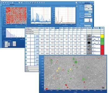
Screenshots from LabSpec 6 showing located particles captured white light optical image together with statistical table and spectra acquisition. Image Credit: HORIBA
How it Works
The ParticleFinder workflow is quite simple and intuitive. The module is completely connected to associated modules for data gathering, processing, analysis, and presentation and is a built-in part of the robust LabSpec 6 software.
Particle Location
Simple processing of the sample optical image allows for the rapid location of particles, creating a binary image that clearly highlights the particle positions.
Full morphological filtering tools are also included to ensure truly meaningful location. The optical image can cover any user-specified area using the integrated video montage/mosaic tool, allowing areas as small as a millimeter or centimeter to be covered.
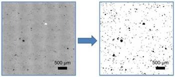
Image Credit: HORIBA
Statistical Analysis
Once the particles have been found, their sizes and shapes, along with statistical analysis, are determined for each particle. The parameters’ histograms (including those for area, perimeter, axis, ellipse ratio, circularity, and brightness) are supplied for simple visualization.
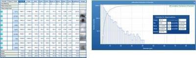
Image Credit: HORIBA
Particle Selection
Initially, all particles located are put up for Raman analysis. They can still be filtered based on the statistical findings, enabling only particles of a given size/shape to be studied. Only particles within a specified size range, for example, can be marked for Raman characterization.
Raman Acquisition
The Raman acquisition stage makes use of all of the Raman instrument’s features, including spectral resolution, confocality, mapping, and laser wavelength selection. These adaptable experiments make it simple to get optimum findings from various sample sets. Along with the statistical characteristics, the Raman spectra are shown in a table.
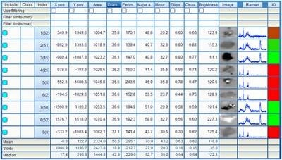
Image Credit: HORIBA
Data Analysis
The individual particles in the binary image are automatically connected to the Raman data after it has been acquired. The spectra can now be processed, examined, and presented using all of LabSpec 6’s robust functionality.
Spectra can be exported to the HORIBA Edition KnowItAll® database searching tool for advanced spectrum matching to known materials.
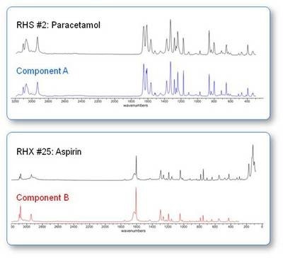
Image Credit: HORIBA
As an alternative, the spectra can be automatically clustered and decomposed using the fully integrated Multivariate Analysis module. Each color will represent a separate component as the multivariate analysis results are shown in the statistical table and directly on the video image of the sample.

Image Credit: HORIBA
Particle Analysis with HORIBA
The ParticleFinder module for Raman spectrometers is part of HORIBA’s expertise in particle analysis.
HORIBA offers powerful, hassle-free tools for particle size, particle shape, zeta potential, and surface area analysis. The measurable particle size ranges from below 1 nanometer to 30 millimeters at concentrations ranging from 1 ppm to 50 vol% with shape determination available starting at 1 micrometer.
A range of technologies is employed, including laser diffraction, dynamic light scattering, and dynamic and static image analysis.
Specifications
- Spectrum export tool for KnowItAll® database searching tool
- Advanced particle clustering and decomposition methods with multivariate analysis
- Full capabilities of the “simply powerful” LabSpec 6 from HORIBA, including full data processing, analysis, and display capabilities
- Automatic location of particles
- Video montage/mosaic tool for large-area analysis
- Characterization of particle size and particle shape
- Select particles according to particle size and particle shape
- Automatic acquisition of Raman spectrum for each particle