The XGT-9000 X-Ray Analytical Microscope (Micro-XRF) represents the next generation of micro-X-Ray fluorescence spectroscopy capabilities in terms of speed and versatility.
- High sensitivity and a broad spectrum of observable elements
- Dual detector types for fluorescent and transmission X-Rays
- ≥15 µm spot size with an ultra-high intensity capillary
- High-resolution cameras and multiple illumination modes
- Flexible and user-friendly software interface
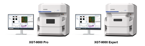
Image Credit: HORIBA Scientific
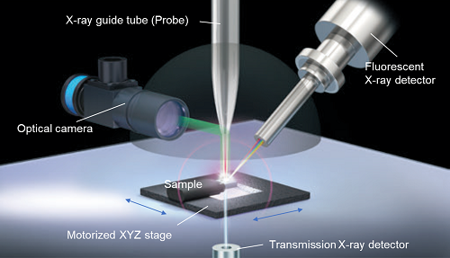
Image Credit: HORIBA Scientific
XGT-9000
Video Credit: HORIBA Scientific
Features
Higher sensitivity and wider detection range by new detection systems
The XGT-9000 Pro has improved sensitivity thanks to innovative detection methods, while the XGT-9000 Expert has the highest sensitivity and the widest detectable element range. Increased sensitivity reduces measurement time and increases labor efficiency, while a larger detectable element range broadens application options.
![(Left) Cu intensity comparison (Right) Super light elements intensity comparison.[1][2] All the results are compared with XGT-9000 Pro/Expert vs. HORIBA conventional micro-XRF models.](https://www.azom.com/images/equipments/EquipmentImage_8546_16819827050168071.png)
(Left) Cu intensity comparison (Right) Super light elements intensity comparison.[1][2] All the results are compared with XGT-9000 Pro/Expert vs. HORIBA conventional micro-XRF models. Image Credit: HORIBA Scientific
High-quality macro and micro imaging with multiple probe selection
The XGT-9000 Series offers maximum performance and versatility due to its well-designed excitation system, which includes a high-output X-Ray generator (up to 50 kV and 1000 µA) and a wide range of probe spot sizes ranging from 10 microns to 1.2 mm. Multi-probes can be inserted into the instrument and switched on and off through software.
It permits quick, high-quality imaging at the macro and microscopic levels without sacrificing the need for spatial resolution and measurement time over a broad map area. There are two ultra-high intensity probes available: 15 µm and 100 µm.
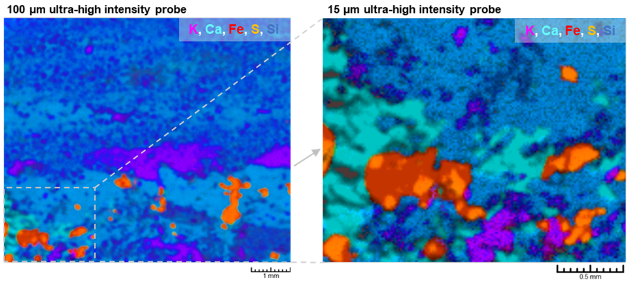
Elemental layered images on a Lapis lazuli stone using multi-probes (Left) Fast scanning using 100 µm ultra-high intensity probe. (Right) Detailed imaging using 15 µm ultra-high intensity probe. Image Credit: HORIBA Scientific
Dual types of detectors for fluorescent X-Rays and transmission X-Rays
The XGT-9000 Series offers dual types of detectors. One is a fluorescent X-Ray detector that reveals a sample’s elemental distribution, and the other is a transmission X-Ray detector that reveals a sample’s interior structure.
The XGT-9000 can concurrently acquire images of the two different types of the same region. It is beneficial for a deeper comprehension of electronics, gemstones with inclusions, and biological specimens.
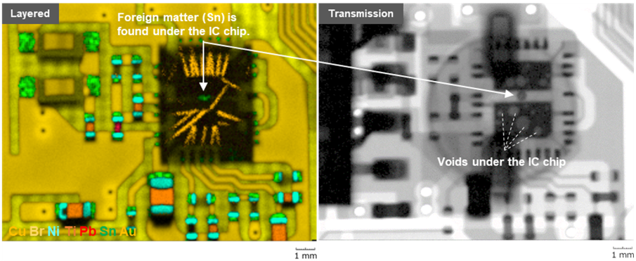
Simultaneous imaging of elemental image and transmission X-ray image of a printed circuit board (Left) Elemental layered image revealed a foreign matter stuck under the IC chip. (Right) Transmission X-ray image revealed many voids under the IC chip. Image Credit: HORIBA Scientific
High resolution and brilliant optical images
For micro-XRF analysis, crystal-clear images must be captured. The XGT-9000 series has high-resolution cameras to capture an image of the entire sample and its features down to the microscopic level.
Both images can be digitally zoomed in and out to locate the ideal place to examine on a sample surface with flexibility. Various illumination systems allow users to obtain stunning optical images of the target sample.
| (Left) Whole image |
(Right) Detailed image |
 |
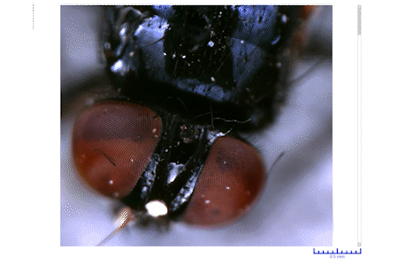 |
High resolution and brilliant optical image of a fly. Image Credit: HORIBA Scientific
Wide applications of the sample chamber
Micro-size fragments, printed circuit boards, coins, sheets, powder, liquid, and wafers are just a few samples that can be used with the XGT-9000 Series. Sample holders come in a variety of designs. Sensitivity can be pulled up by selecting one of four measurement environments.
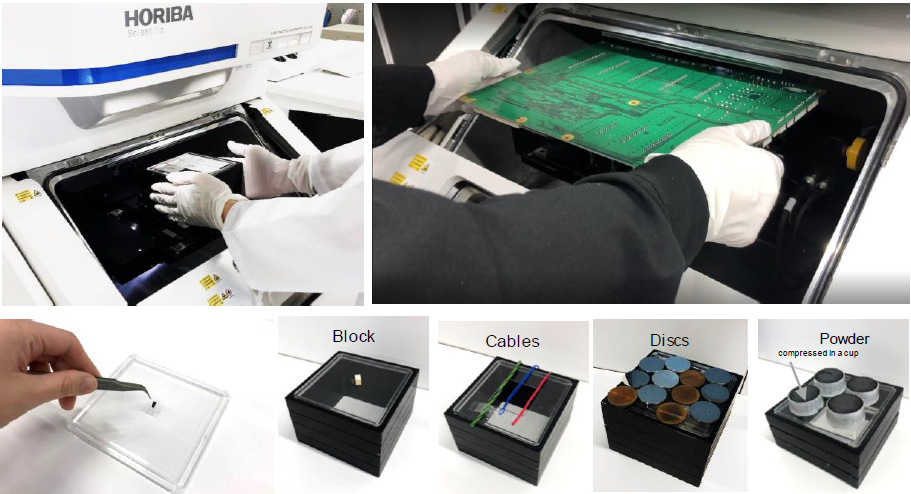
Wide applications of the sample chamber. Image Credit: HORIBA Scientific
Flexible and user-friendly software
The user-friendly interface of the XGT-9000 software is highly flexible. At a glance, it shows the periodic table, the data tree, optical images, spectrum results, and map imaging results.
Users can alter the screen’s layout with flexibility. The outcomes can also be shown on several monitors. It improves users’ data analysis and lets them view the results more clearly.
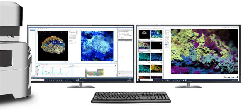
An example of software layout on dual monitors. Image Credit: HORIBA Scientific
Advanced modules and regular software features can be added to the software suite to offer users a more comprehensive user experience.
- Multilayer FPM module for thickness measurement with or without standard specimens
- RoHS module for RoHS screening
- A queue module for automated multiple measurements in unattended mode
- Particle Finding module for particle analysis and co-localized analysis
- LabSpec Link module for data transfer to LabSpec 6 for multivariate analysis
Application Examples
- HORIBA’s contributions to the exploration and analysis of the Ryugu asteroid
- Tracing toxic heavy metals in organisms using X-Ray fluorescence (XRF)
- Raman and X-Ray fluorescence spectroscopies reveal the mysteries behind the ancient Japanese art form
- Mars life signs probed by spectroscopy
- How XRF may help uncover hidden clues to life on Mars

Image Credit: HORIBA Scientific
Specifications
Source: HORIBA Scientific
| Model |
XGT-9000 |
| XGT-9000 Pro |
XGT-9000 C |
XGT-9000 Expert |
| Basic information |
| Instrument |
X-Ray analytical microscope |
| Principle |
Energy dispersive X-Ray fluorescence spectroscopy |
| Detectable elements* |
F (9) - Am (95) |
C (6) - Am (95) |
B (5) - Am (95) |
| Available chamber size |
450 mm (W) x 500 mm (D) x 80 mm (H) |
| Maximum mass of sample |
1 kg |
| Maximum mapping area |
100 mm x 100 mm on 300 mm (W) x 250 mm (D) |
| Sample observation |
| Optical image observation |
Two high-resolution cameras |
Whole image
Detailed image |
5 million pixels, Field of view: 100 mm x 100 mm
5 million pixels, Field of view: 2.5 mm x 2.5 mm |
| Optical design |
Vertical-coaxial X-ray and optical observation |
| Sample illumination / Observation |
Top, bottom, side illuminations / Bright and dark fields |
| X-Ray generator |
| Power |
Up to 50 W |
| Voltage |
Up to 50 kV |
| Current |
Up to 1 mA |
| Target material |
Rh |
| X-Ray guide tube (Probe) |
| Probe spot size selection |
Various probe combination can be offered
(e.g. 15 μm ultra-high intensity probe and 100 μm ultra-high intensity probe can be chosen) |
| Detectors |
| X-ray fluorescence detector |
Liquid nitrogen-free Silicon Drift Detector (SDD) |
| Transmission detector |
NaI (Tl) |
| Operating mode |
| Measurement environment |
Whole vacuum
Partial vacuum
Whole ambient
He purge (optional) |
Whole vacuum
Partial vacuum
Whole ambient
He purge (optional) |
Whole vacuum
Partial vacuum |
| Instrument dimension (main unit) |
| Instrument size |
680 mm (W) x 860 mm (D) x 760 mm (H) |
| Mass weight |
Approximately 200 kg |
*Under whole vacuum condition
Dimensions (unit: mm)
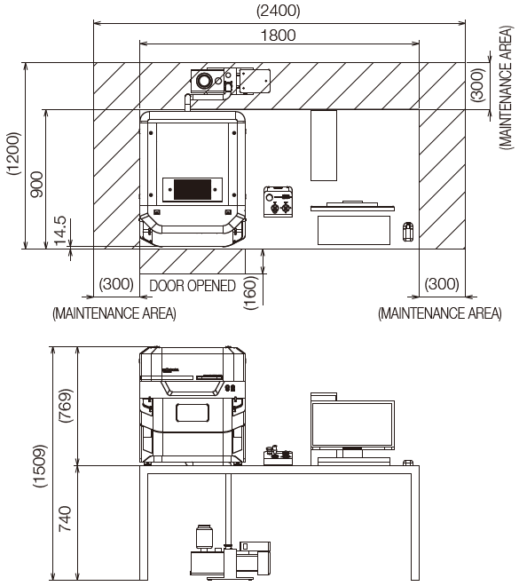
Image Credit: HORIBA Scientific
Applications
Proton exchange membrane fuel cell material analyses using micro-XRF
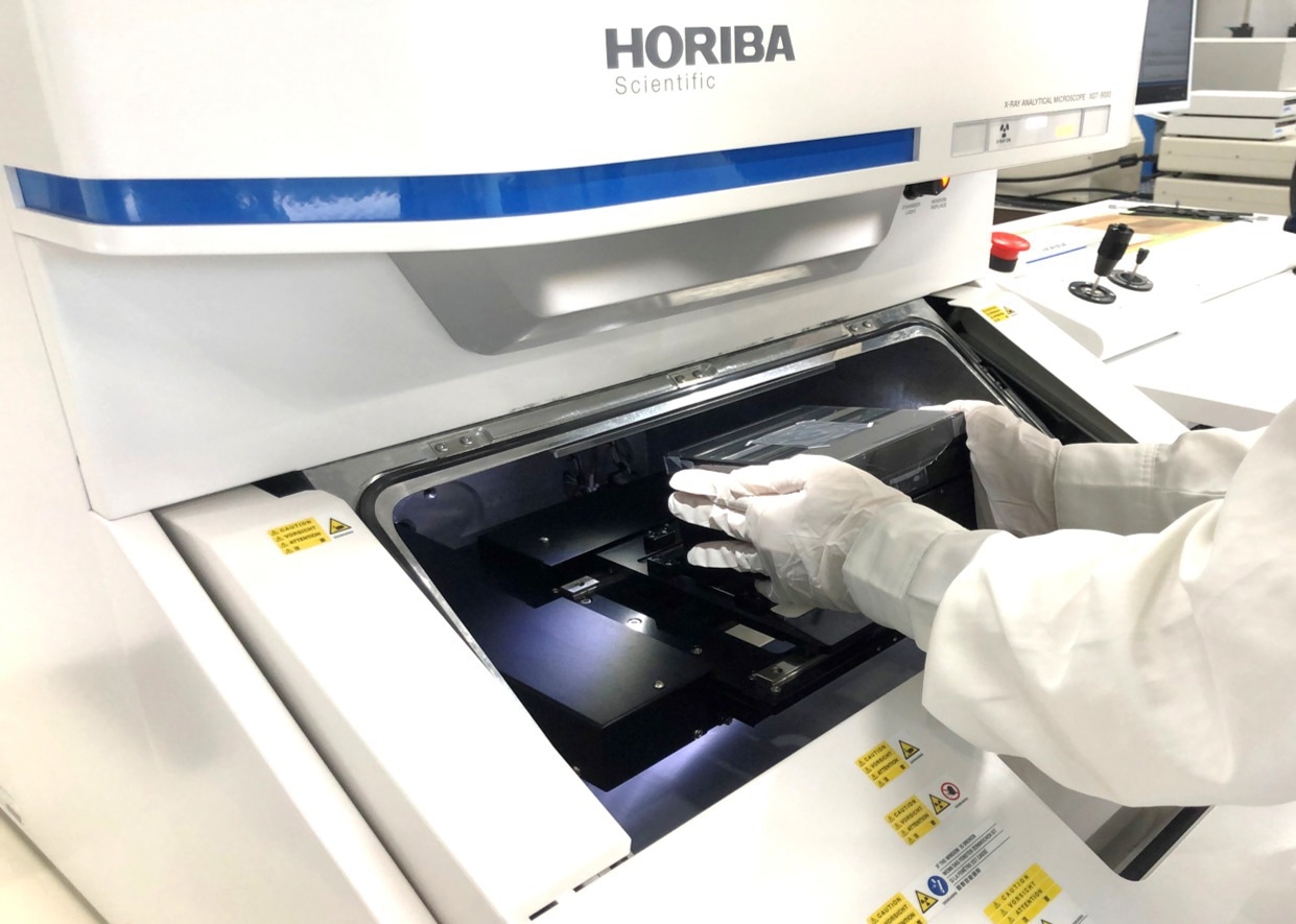
Image Credit: HORIBA Scientific
Proton exchange membrane fuel cell (PEMFC) is made up mostly of organic materials, such as proton exchange membranes, carbon supports, and carbon sheets. However, it also contains inorganic materials, such as radical quenchers and precious metal catalysts.
Glass-filled ruby characterization with the XGT-9000, HORIBA’s new Micro-XRF
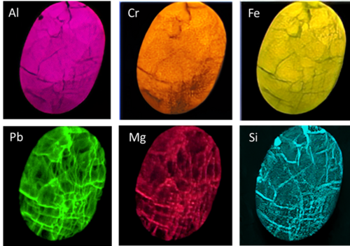
Image Credit: HORIBA Scientific
Gemstones are heated, irradiated, or have cracks filled to make them perfect for jewelry. A ruby provided by Mineralab was imaged using an XGT-9000 X-Ray analytical microscope.
The ruby was found to have multiple defects using transmission X-Ray imaging, and fluorescent X-Ray imaging showed that the ruby surface had been filled with Pb, Mg, and Si, representing lead glass.
Non-destructive thickness and composition analysis of NiP/Au plating on Cu contacts using micro-XRF
Image Credit: HORIBA Scientific
Using a HORIBA X-Ray analytical microscope, plating thickness and composition were analyzed on dual-layer NiP/Au plating on Cu contacts on a flexible printed circuit.
Without any sample preparation, Au of an ultra-thin layer was effectively detected. The thickness findings of the Au and NiP plating were reliable and consistent with the given values.
Pigment identification of an old Japanese flag “Hinomaru” using micro-XRF and Raman microscopy
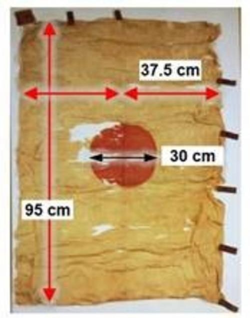
Image Credit: HORIBA Scientific
Here, pigment identification using micro-XRF and Raman microscopy on a Japanese flag called “Hinomaru,” which is thought to be the oldest and was created by an ancient Japanese Emperor named Go-Daigo, is presented.
The two spectroscopic studies revealed that the cinnabar ore (Mercury (II) sulfide) extracted from the Mine near the Emperor’s residence matched the red pigment on the flag.
Spectroscopic analysis explains the mystery of Dragonfly eye beads
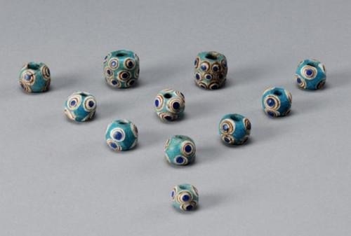
Image Credit: HORIBA Scientific
Spectroscopic investigation can identify the origins of cultural heritages and their historical context.
The bead was identified as being from the Eastern Mediterranean region using Raman spectroscopy and X-Ray analytical microscopy. The finding revealed China had cultural and economic exchanges with them at the time.
Optical micro-spectroscopies on a path to identify the source of life
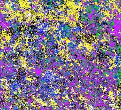
Image Credit: HORIBA Scientific
Combining Raman and X-Ray Fluorescence microscopies can provide insight into the cosmos' birth.
Non-destructive failure analysis on electronic components using the XGT-9000
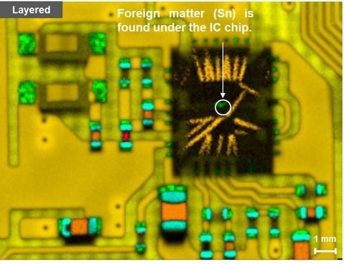
Image Credit: HORIBA Scientific
Since X-Rays have a high penetration rate, μ-XRF is a non-destructive analytical technique that can examine flaws in the material, even those that are invisible.
The XGT-9000’s major characteristics, including vertical irradiation of a 10 μm probe and simultaneous imaging of fluorescent and transmission X-Rays, are used in a failure analysis to identify voids, foreign materials, and ion migration on electronics.
Multiple characterizations of a blister pack using the XGT-9000
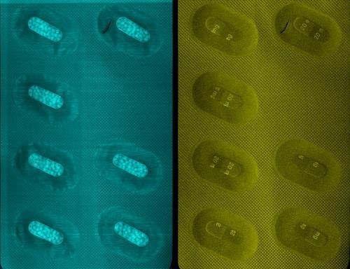
Image Credit: HORIBA Scientific
The XGT-9000 is a new X-Ray microscope by HORIBA. Its analytical flexibility enables a variety of characterizations to be carried out on a blister pack, from full-pack mapping to assessments of the particle size of the capsule content.
Characterization of pyrite inclusions in lapis lazuli using X-Ray fluorescence micro-imaging
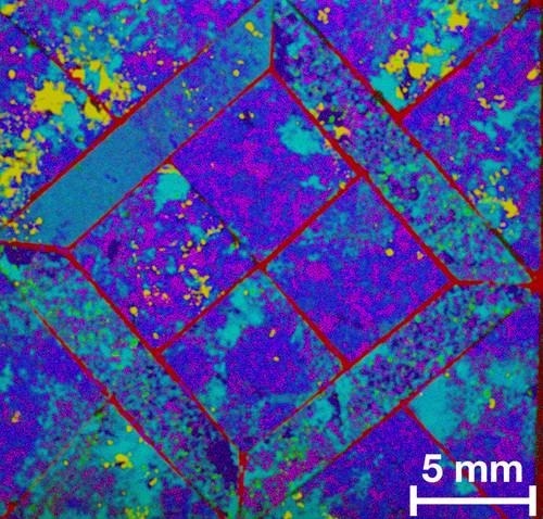
Image Credit: HORIBA Scientific
A deep blue metamorphic rock called lapis lazuli is used as a semiprecious stone, however, it has inclusions that can lower the price. The XGT-9000, a new X-Ray microscope from HORIBA, is used to examine the distribution of main elements and pyrite impurities.
Elemental distribution imaging on edible insects using micro-XRF

Image Credit: HORIBA Scientific
Due to their nutritional advantages, such as their protein, fat, and mineral content, edible insects have drawn interest as a potential option to help eliminate food insecurity. Without causing any damage, micro-XRF can be utilized to comprehend the elemental distributions in insects.
Using a HORIBA XGT-9000 X-Ray analytical microscope, HORIBA Scientific conducted elemental map imaging on edible crickets, revealing a rich supply of zinc in their jaws.
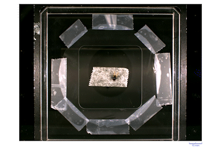
Foreign matter analysis in food using the XGT 9000
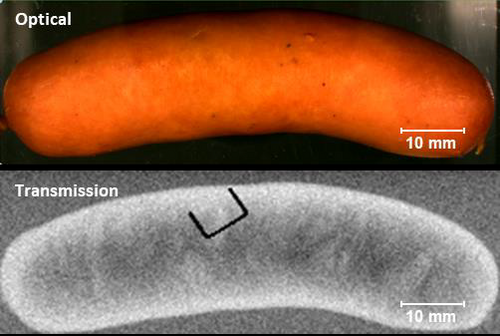
Image Credit: HORIBA Scientific
Here is an example of a foreign matter analysis in food items utilizing the XGT 9000. The following three analyses are available: A fly was discovered in a drink product, along with foreign particles on an oily salami and foreign material within laminated ham and sausage.
The non-destructive identification of black ink in a tempered document using XGT-9000
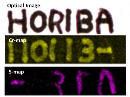
Image Credit: HORIBA Scientific
One of the effective non-destructive analytical methods used in forensic science is μ-XRF. The XGT-9000’s elemental mapping and spectrum search capabilities make it possible to detect purposeful alternation and determine the type of ink that was used on a document.
QC of semiconductors which feature thin and narrow patterns
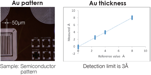
Image Credit: HORIBA Scientific
The XGT-9000 is a practical instrument for semiconductor quality control since it combines microbeam and thickness measurement capabilities. They feature thin and narrow patterns. The thickness sensitivity can be at the Angstrom level and relies on the elements being traced.
QC, counterfeit products, presence of foreign materials
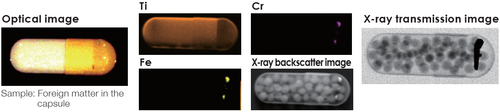
Image Credit: HORIBA Scientific
The material that is enclosed can partially absorb X-Ray fluorescence photons, which means they will not be seen in the spectrum. The full view is seen in the X-Ray transmission image.
Foreign particle Analysis on a Separator Film of Lithium-ion Battery
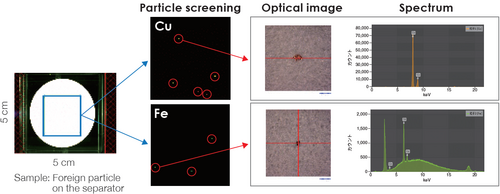
Image Credit: HORIBA Scientific
The XGT-9000 can locate the source of contamination by detecting foreign particles and analyzing their composition.
Failure analysis, RoHS testing of electronics
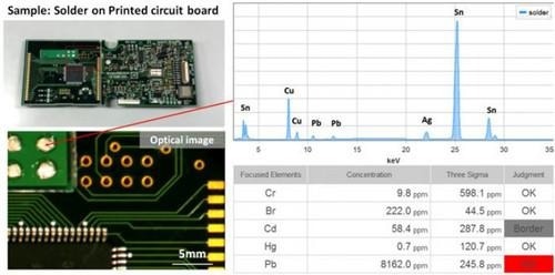
Image Credit: HORIBA Scientific
To detect flaws inside electronic components, simultaneous imaging of transmission and fluorescent X-Rays is beneficial.
Surface analysis of corrosion/contamination for material science
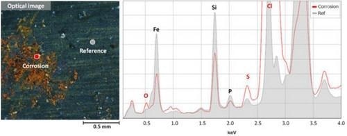
Image Credit: HORIBA Scientific
For material science, elemental distribution is essential. This information is presented non-destructively via the XGT-9000 Series.
Elemental composition identification of geoscience/mineralogical samples
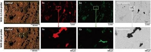
Image Credit: HORIBA Scientific
The XGT-9000 Series’ wide range of probes and spot sizes allows for thorough and in-depth analysis of geological and mineral samples.
Trace evidence identification, fake product identification of forensic samples
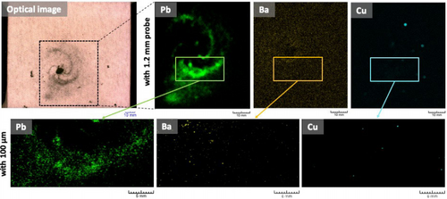
Image Credit: HORIBA Scientific
The XGT-9000 Series can be used to identify traces of evidence, including fibers with diameters as small as tens of microns, fragments of glass, and residue from gunshot wounds that have been collected.
Metabolism investigation of biological samples
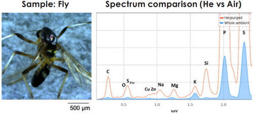
Image Credit: HORIBA Scientific
The XGT-9000 Series can be utilized to identify traces of evidence, including fibers and glass fragments as small as tens of microns in size, gunshot residues, and collected residues.
Gun shot residue analysis using X-Ray fluorescence micro-analysis
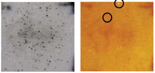
Image Credit: HORIBA Scientific
Individual minute particles can be identified for their elemental composition using a micro-XRF analysis of gunshot residue. Automated element imaging delivers element distribution maps with great spatial resolution, enabling precise analysis of particle sizes and shapes.
X-Ray micro-analysis for pearl characterization in forensic science
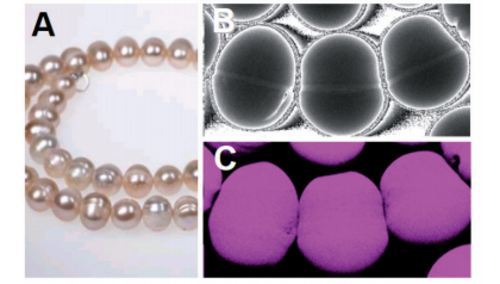
Image Credit: HORIBA Scientific
The composition and structure of pearls can be better understood by using simultaneous XRF and transmission X-Ray imaging. Customs officers need this information so they can easily evaluate whether pearls are cultured, natural, or fake.
Fingerprint imaging with micro-XRF
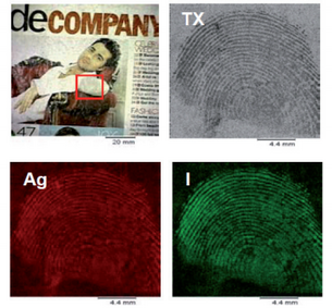
Image Credit: HORIBA Scientific
High spatial resolution elemental mapping studies offer a helpful approach to fingerprint analysis in circumstances where more conventional techniques fail. It has been chemically processed and then imaged to create fingerprints on glossy paper and finely woven fabrics.
Micro-XRF analysis for lead contamination in toys

Image Credit: HORIBA Scientific
A plastic toy’s numerous parts are examined for the presence of lead. Spot analysis reveals that levels of this dangerous substance can rise to 0.3%. Its distribution throughout the toy could be immediately identified via XRF imaging.
Fast thickness measurement of thin metal coatings by Micro-XRF
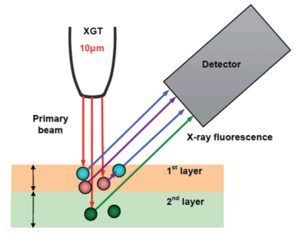
Image Credit: HORIBA Scientific
With just one measurement, multi-layered materials can be characterized due to the EDXRF analysis’s penetrating nature. Even minute structures, such as bonding pads on circuit boards, could have their composition and layer thickness examined with great spatial resolution.
Quality control and defect analysis in the electronics industry using Micro-XRF
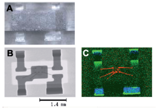
Image Credit: HORIBA Scientific
On both individual components and complete circuit boards, troubleshooting and defect evaluations of components buried within opaque resins are addressed. For assuring compliance with the WEEE/RoHS requirements, quantitative analysis down to the ppm level is desirable.
Micro-XRF analysis for the electronics industry
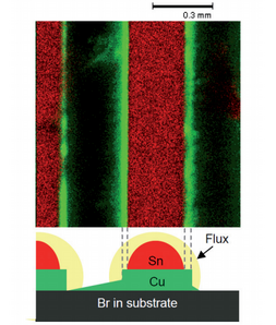
Image Credit: HORIBA Scientific
The XGT-5000 is the instrument of choice for quick examination of electronic components, whether for analysis of banned dangerous elements (the WEEE/RoHS “lead-free” regulation), troubleshooting, or R&D. This is due to its combination of ground-breaking spatial resolution and sensitivity.
Elemental analysis of single rice grains using XRF micro-analysis
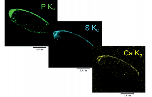
Image Credit: HORIBA Scientific
To investigate the impact of grain polishing, individual rice grains are subjected to micro-XRF analysis. The amount of polish a grain has can be correlated with the number of mineral elements present.
Elemental micro-analysis of leaves using EDXRF
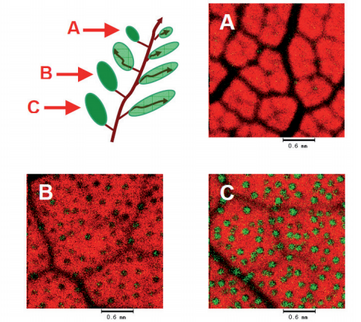
Image Credit: HORIBA Scientific
Mulberry leaves are found to have calcium micro-nodules, and XRF-mapped imaging shows a relationship between nodule concentration and leaf age. The absorption of heavy element pollution by plants is examined in separate research, and high-resolution images of the distribution of lead through the leaf are swiftly acquired.
Biological applications of XRF microscopy
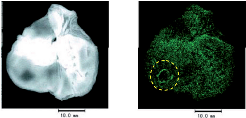
Image Credit: HORIBA Scientific
Researchers have looked at how zinc affects the healing of stomach ulcers; mapped imaging of tissue samples reveals signs of zinc buildup within the ulcerated tissue.
A fish otolith, or “ear bone,” has been examined in different research to reveal its complex physical structure and heterogeneous elemental composition.
Micro-XRF for non-Destructive analysis of museum and archaeological objects
Image Credit: HORIBA Scientific
An ancient glass burial ornament has been examined to determine the particular coloring additives employed, and the pigments used in an old Nepalese book have been analyzed and assigned.