The Partica Particle Size and Shape Analyzer combines laser diffraction with dynamic image analysis for quicker, more thorough particle analysis.
Partica integrates these complementary methods into one device, enabling the simultaneous capture of particle size distribution and shape, thereby minimizing measurement duration, optimizing R&D and QC processes, and conserving laboratory space.
Key Features
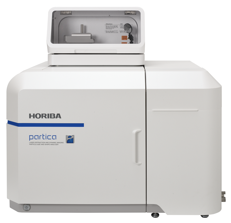
Image Credit: HORIBA
- The all-in-one system integrates laser diffraction and scattering for measuring particle size distribution, along with dynamic image analysis for particle shape analysis.
- Facilitates the analysis of particle size and shape for various sample types, including liquids, slurries, and powders.
- The two-camera setup allows for particle imaging over a broad size spectrum.
- Highly customizable and scalable, designed to maximize expandability.
Features
Laser Diffraction and Dynamic Image Analysis Particle Size and Shape Analyzer: Two Systems in One
- Facilitates the analysis of particle size and shape across a wide variety of sample types and concentrations, ranging from water-soluble substances like pharmaceuticals and food products to dry powders and high-solids samples, including battery materials.
- By integrating both techniques into one cohesive system, Partica greatly minimizes overall measurement duration, enhances efficiency in research and development as well as quality control, all within a notably smaller footprint.
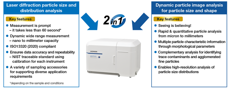
Image Credit: HORIBA
Particle Size and Shape Analysis for a Wide Range of Sample Forms, Including Liquids, Slurries, and Powders
- Partica's laser diffraction technology boasts industry-leading precision (within ±0.6% of verified reference particle standards) and a broad measuring range (10 nm to 5 mm). Further modifications to the optical system result in greater sensitivity.
- High-resolution optics record precise particle shape information from approximately 1 µm to the millimeter scale for dynamic image analysis.
- More than thirty particle shape metrics, such as equivalent diameter, minimum/maximum (Feret) diameter, circularity, and aspect ratio, are extracted by the system for shape analysis.
Silica Slurry Quality Analysis
Using a variety of technologies improves the study of particle size and distribution, providing a more comprehensive picture of the material testing. This is an illustration of how static light scattering and imaging technologies can be used to enhance one technique.
Comparing the two methods, this silica slurry analysis shows very few large impurities or agglomerated particles while offering comprehensive characterization data that can assist in identifying and resolving possible problems throughout the production quality control process.
Partica's simultaneous dual-mode analysis combines laser diffraction measurement with particle imaging, providing complementary results in a single test.
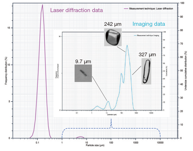
Image Credit: HORIBA
Two-Camera System Captures Particle Images Across a Broad Range
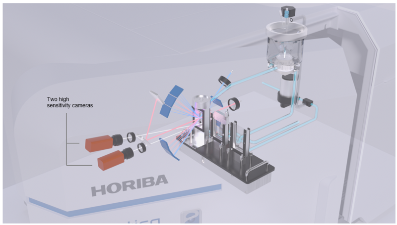
Image Credit: HORIBA
- The device allows for a wide range of measurements, from around 1 µm to several millimeters, by combining two cameras with varying resolutions.
- It captures up to 300 frames per second at peak, enabling analysis of tens of thousands of particles in a short time.
- This approach removes the necessity for lengthy hardware modifications, such as changing microscope objectives or swapping apertures, which are typically required in methods based on Coulter counters.
Specifications
Source: HORIBA
| Model |
|
LA-1000 series |
LA-970 series |
| Measurement principle |
DIA |
Dynamic imagE analysis |
- |
| LD |
Mie scattering and Fraunhofer diffraction |
| Total measurement range |
|
0.01–8000 μm |
0.01–5000 μm |
| Measurement range (Wet) |
DIA |
0.3–3000 μm* |
- |
| LD |
0.01–3000 μm |
| Measurement range (Dry) |
DIA |
1.1–8000 μm* |
- |
| LD |
0.1–5000 μm |
| Light sources |
|
LD (650 nm): 5 mW, LED (405 nm): Maximum 10 mW |
| Number of cameras |
|
Two (dual-camera system) |
- |
| Camera performance |
|
Maximum 300 fps |
- |
| Digital resolution |
|
Minimum 0.3 μm |
- |
| Measurement parameters |
|
More than 30 image analysis parameters |
- |
| Dimensions and weight ** |
|
720 mm (W) x 565 mm (D) x 470 mm (H): approximately 56 kg |
| Optional accessories |
|
Powderjet Dry Powder Feeder, MiniFlow unit, Fraction cell, High Concentration cell, Paste cell |
* Depends on particle shapes
** Dimensions and weight exclude pipework and other protrusions, and optional accessories.
Optional Accessories
Source: HORIBA
| . |
. |
. |
MiniFlow (Circulation
system) |
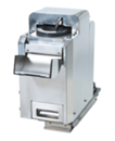 |
Small-volume circulation with organic solvents.
- Measurement needs as little as 35 mL of solvent (180 mL with conventional circulation systems)
- Dispersion is possible with the built-in ultrasonic probe
|
| Dry Powder Feeder |
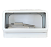 |
- Can measure samples in a powder state
- Supports both non-dispersed measurement by free fall and forced-dispersion measurement using compressed air
|
High Concentration
Cell* |
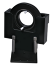 |
The High Concentration Cell can measure close to the original concentration with low dilution or without dilution.
- Measure inks, paints, pigments, and emulsions without dilution
- Positive and negative electrode materials of secondary batteries, etc.
- Observe changes in the agglomeration state based on concentration, etc.
- Agglomeration was found when a battery was measured near the original concentration
|
| Paste Cell* |
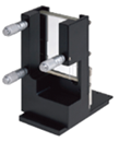 |
The Paste Cell can measure undiluted samples or samples dispersed in a viscous medium.
- Microparticles dispersed in high-viscosity samples or polymers
- Measure magnetic powder by dispersing it in viscous oil to prevent re-agglomeration
|
| Fraction Cell |
|
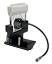
The Fraction Cell can take measurements with samples as small as only 5 mL, minimizing the amount of dispersant. The Fraction Cell is ideal when the sample is extremely small, or an entire sample needs to be recovered.
Measurement range: 0.01–1000 µm, suits organic solvents
Cell volumes: 5 mL, 10 mL, 15 mL
- Rare samples
- Measurements with highly volatile solvent
|
*These optional accessories are available for laser diffraction analysis.