The JSM-IT210 InTouchScope™ SEM Series offers advanced JEOL technology and automation in a compact design.
Smart – Flexible - Powerful
Smart
The InTouchScope™ series SEMs are now more accessible to all levels, thanks to recent developments. A Specimen Exchange mode walks a new operator through the process of introducing samples, automatically adjusting conditions, and creating images. JEOL's intelligent technology provides best-in-class auto functions from alignment to focus, resulting in fast, clear, and excellent images.
The built-in optical camera for navigation and seamless transition to SEM imaging makes the operation quick and simple. Our analytical models allow you to view live EDS spectra and X-ray maps. Take it to the next level with built-in automation, including Montage (large area mosaics) and Simple SEM, which collects images automatically at numerous locations, magnifications, and conditions. All of this technology is packed into a small platform, providing remarkable ease of use.
Flexible
Select a platform that is suitable for the user. Both high and low vacuum variants, with or without the Live (integrated) EDS system, are provided. [JSM-IT210, JSM-IT210A, JSM-IT210LV, and JSM-IT210LA].
Powerful
This is a high-resolution tungsten source with unparalleled low-voltage performance. Improved algorithms for automatic beam alignment, autofocus, and stigma correction allow users to concentrate on the results. The high-precision, 5-axis motorized stage increases throughput.
Zeromag’s integrated color camera enables easy navigation to the area of interest and a smooth transition to SEM imaging and analysis. The high-sensitivity quadrant BSE detector allows live 3D surface reconstruction, which improves the view of specimens with complicated topography.
Analytical models include JEOL’s fully embedded EDS system, which provides Real-Time EDS spectra and Live X-ray maps. Internal Data Management connects all data, providing an instant picture of analysis locations.
Key Features
- Specimen Exchange: A comprehensive step-by-step approach from specimen introduction to automatic image creation. Unprecedented ease of use at all levels
- Zeromag: Simplifies navigation by allowing a smooth transition from optical to SEM images. All data is linked, including color images, SEM images, and EDS data for a map of all analysis locations
- Live EDS: Full integration of JEOL EDS with Real-Time Live spectrum and Live X-ray map
- Automation. Montage: Automate large-area image mosaics (stitching) and EDS maps (analytical models)
- Simple SEM: Simplify the workflow and automate routine imaging. Recipes for various kV, detectors, magnifications, and more!
- Signal Depth: This function improves knowledge of analytical spatial resolution by showing the generation depth of distinctive X-ray signals
- There are both high and low vacuum models for any sample type. Easy one-click switch from high to low vacuum
- Easy Maintenance: No specific facilities or compressed gas are required. Plugs into a regular lab wall outlet. Simple source change and automated alignments
Zeromag
Easy transition from optical to live SEM images.
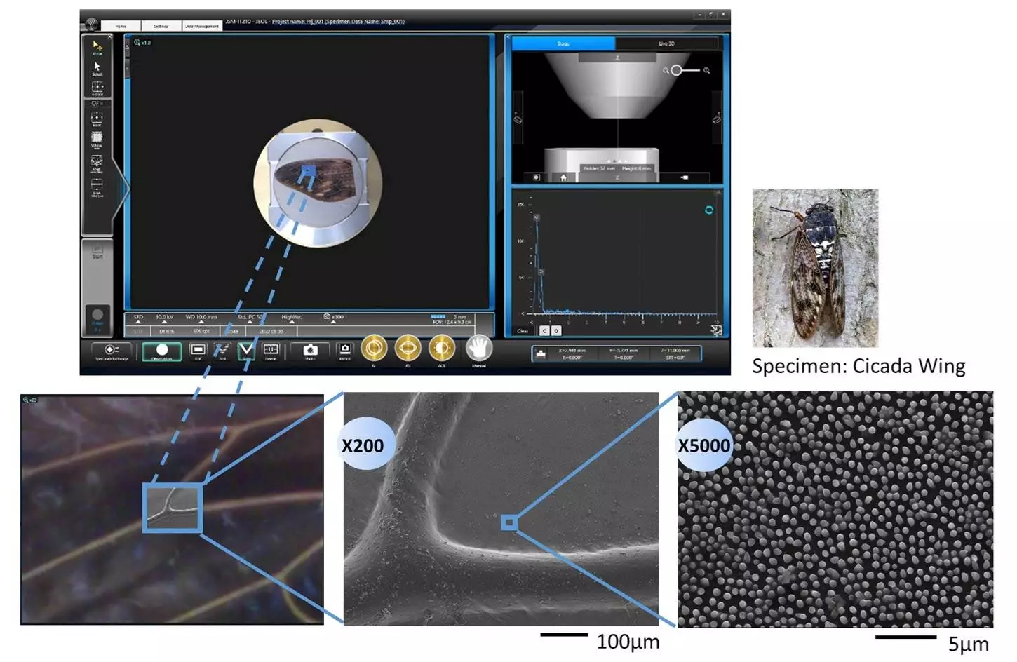
Image Credit: JEOL USA, Inc.
Real-Time EDS – Live Analysis
Embedded JEOL EDS for Live Analysis. View the Live Spectrum or Live Map. Change views with a single click.
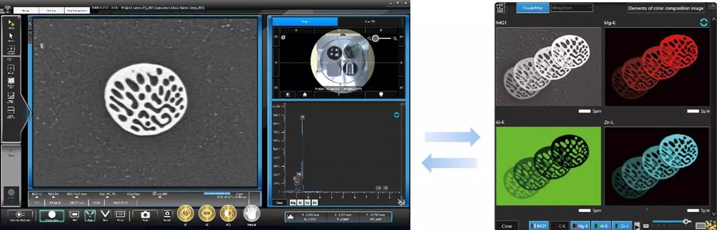
Image Credit: JEOL USA, Inc.
Live 3D
The Quadrant Backscatter Detector can display a live 3D image, offering additional information for samples with complex terrain.
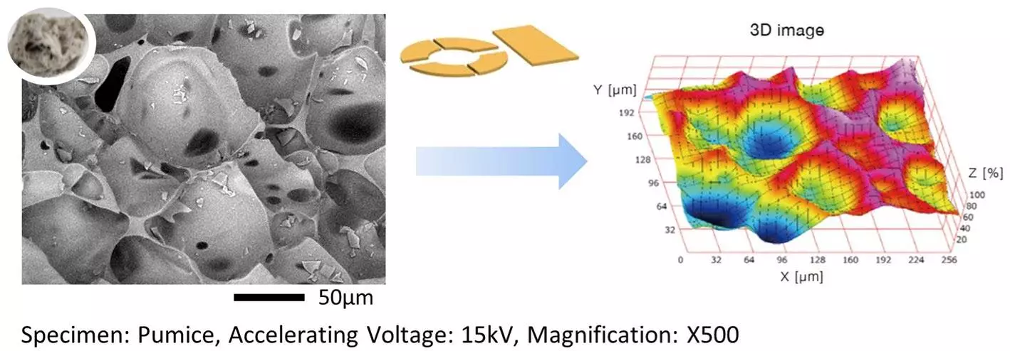
Image Credit: JEOL USA, Inc.
Smile View™ Map - Option
Smile View™ Map offers powerful capabilities for image enhancement, colorization, 3D surface reconstruction, surface metrology, and texture analysis.
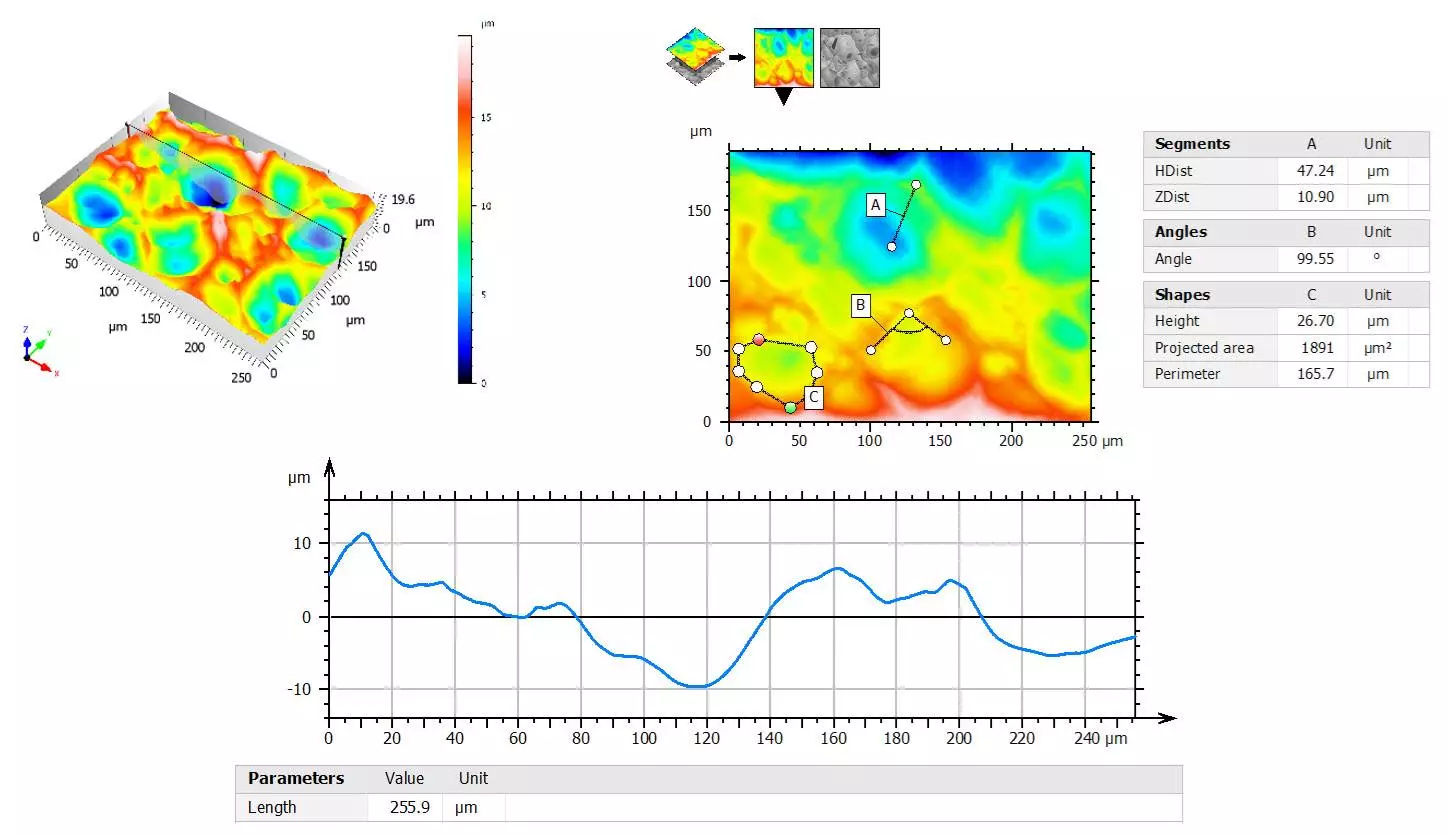
Image Credit: JEOL USA, Inc.
Automation – Montage (Automatic Large Area Image Capture)
Capture images automatically and stitch them together to automatically create a montage image. Includes X-ray maps and analytical models.
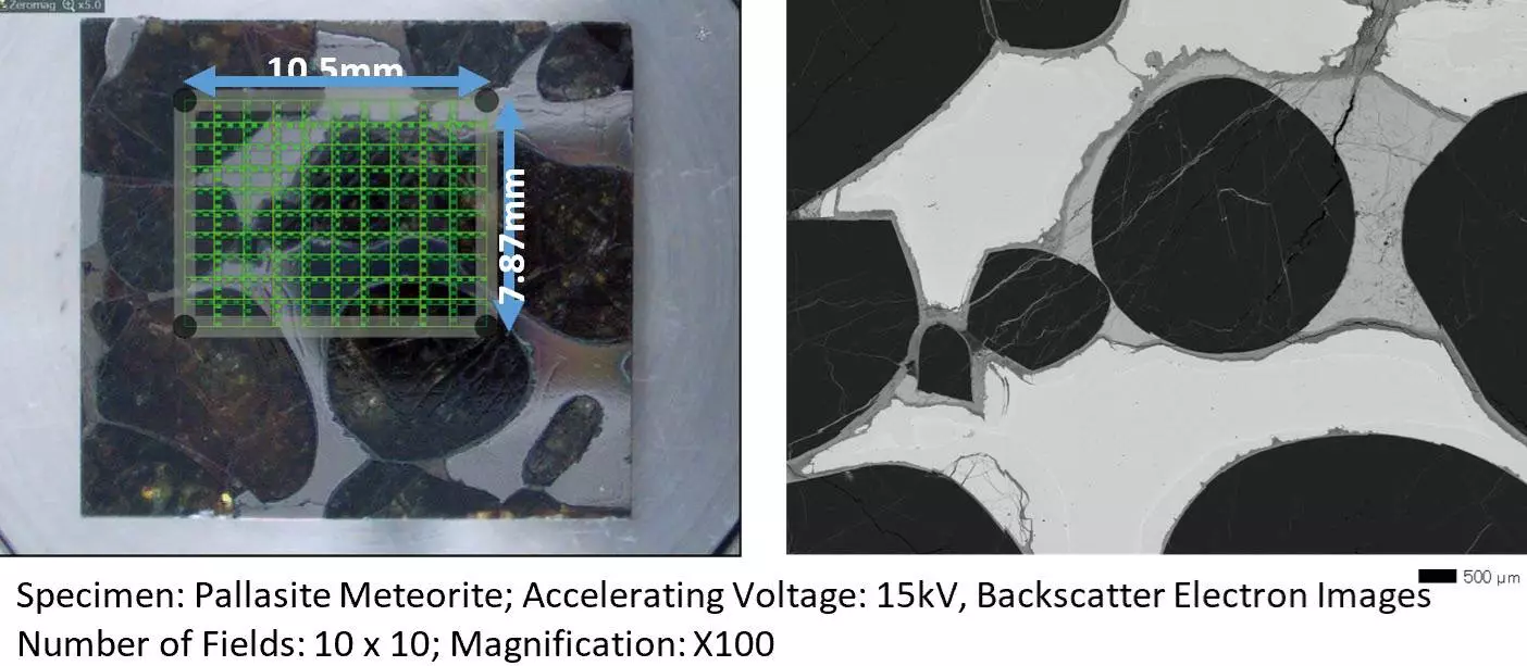
Image Credit: JEOL USA, Inc.
Automation – Simple SEM for Image Capture
Automate image collecting in many places, circumstances, and magnifications:
- Establish Conditions
- Set Target Areas
- Start!
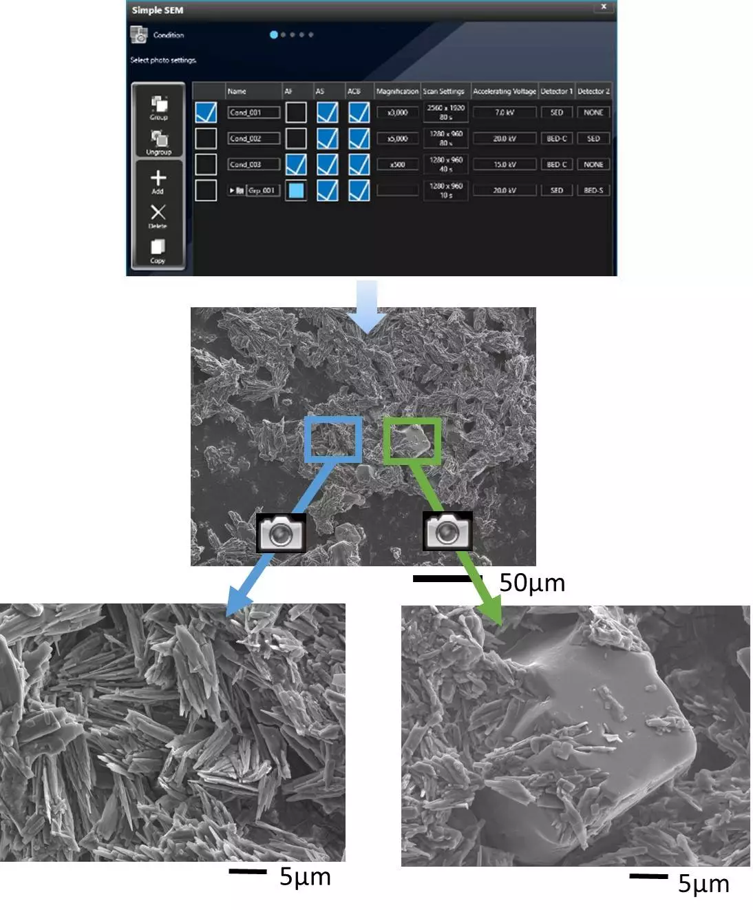
Image Credit: JEOL USA, Inc.
Low Vacuum Hybrid Secondary Electron Detector (LHSED)
This detector captures electron and photon signals, producing a high-signal image with enhanced topographic information. Photon mode gathers and displays a luminescence signal (CL).
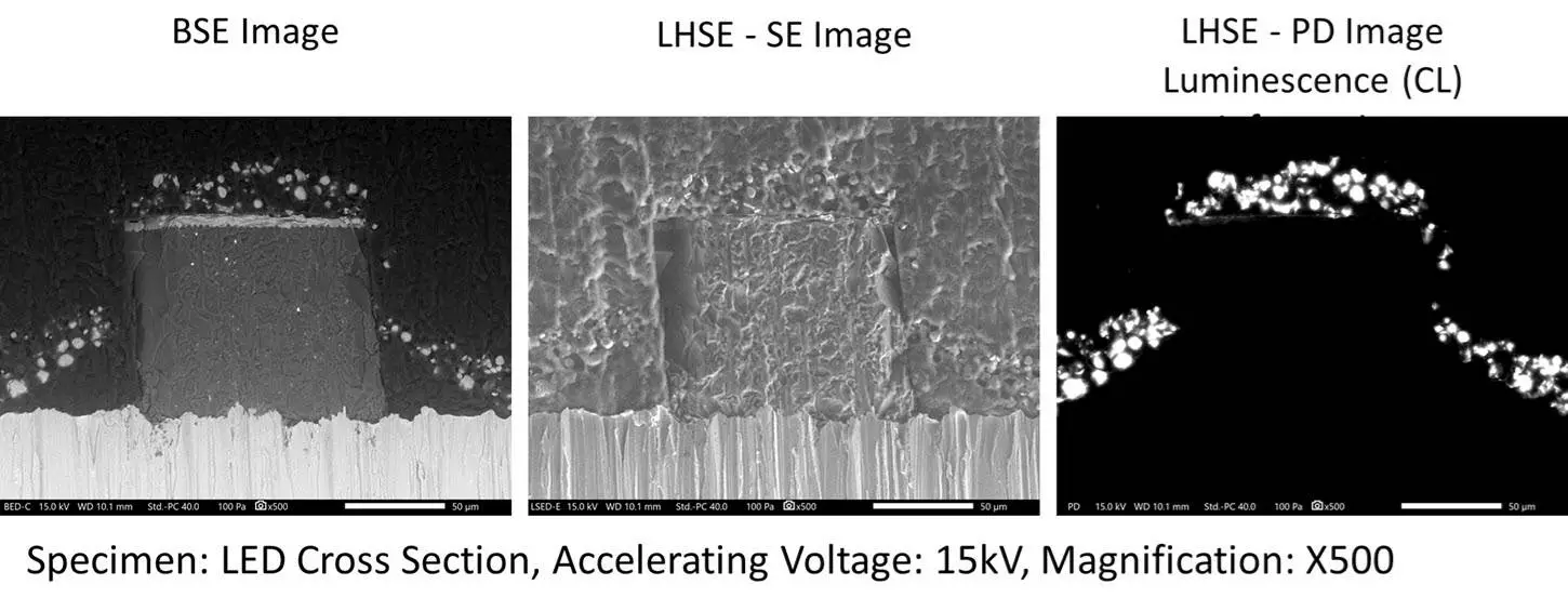
Image Credit: JEOL USA, Inc.