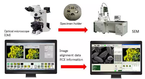
Using a scanning electron microscope and an optical microscope with the same specimen holder is now possible. The system allows researchers to examine fine structures in greater detail by combining optical and electron microscopy.
Using dedicated software, the miXcroscopy™ Linked Optical & Scanning Electron Microscopy System records the exact positions viewed under the optical microscope. It then guides the scanning electron microscope to those same areas for higher magnification and resolution.
The scanning electron microscope can easily observe targets discovered using the optical microscope without having to look for the target again. Images from optical and scanning electron microscopes can be quickly and seamlessly compared and verified.
Key Features
System Outline
Applicable models: Field Emission Scanning Electron Microscope. Electron Probe Microanalyzer (EPMA). Image Credit: JEOL USA, Inc.
Data Acquisition and Intuitive Observation with the Use of Color
The system enhances the SEM image by overlaying colour information from the optical microscope, creating a more intuitive and visually meaningful representation that pure SEM imaging alone cannot provide.
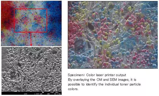
Image Credit: JEOL USA, Inc.
Smooth Target Search Takes Advantage of the Features of the Optical Microscope
Target features can be easily detected using an optical microscope, which are usually difficult to differentiate using SEM images.
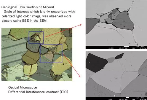
Image Credit: JEOL USA, Inc.
Prevents Damage to the Specimen from the Electron Beam
Using the optical microscope to locate the region of interest helps to avoid contamination or damage from the electron beam. This makes it possible to observe SEM with a low radiation dosage at the observation location.
Applicable models: JXA-8230, JXA-8530F, JXA-8530FPlus. Image Credit: JEOL USA, Inc.
“miXcroscopy™ for EPMA”–Rapid Registration of Analysis Positions
The polarized light microscope, miXcroscopy™ for EPMA, quickly pinpoints analysis positions on specimens where element identification is difficult when using backscattered electron images.
Specimen: Mineral thin section
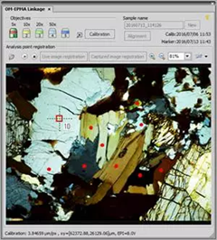
Polarized light OM image: Mag. x50. Image Credit: JEOL USA, Inc.
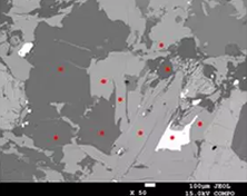
Backscattered electron image: Mag. x50. Image Credit: JEOL USA, Inc.
“miXcroscopy™ for EPMA”–Precise, Fast Analysis of OM-Registered Positions
Conventional Flow
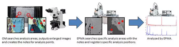
Image Credit: JEOL USA, Inc.
New Flow Using miXcroscopy™ for EPMA
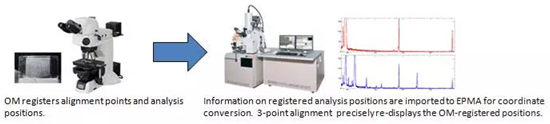
Image Credit: JEOL USA, Inc.
Task separation of OM and EPMA is made more effective with miXcroscopy™ for EPMA. OM records analysis points and searches analysis regions. The elemental analysis is done by EPMA.